
Arrest rhythms
Asystole
No electrical activity in the heart: a flat or nearly flat line, no contraction and no pulse.
Last updated
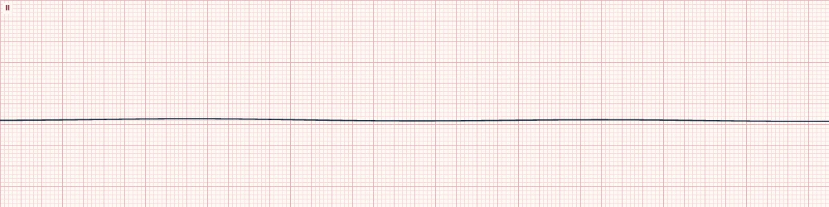
Example tracing, drawn by the lab's engine. Open the lab for a live, adjustable version.
At a glance
- Rate
- None
- Rhythm
- None
- P waves
- None
- QRS
- None
Figures
Still images drawn by the lab's engine at 25 mm/s and 10 mm/mV, with the finding labeled. Select a figure to enlarge it.

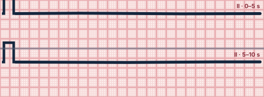
Scroll sideways to see the whole tracing, or select it to enlarge.
What the heart is doing
No pacemaker anywhere in the heart is generating an impulse, so neither atria nor ventricles depolarize. It is usually the end result of prolonged ischemia of the conduction system and muscle.
Why it looks this way
This is asystole because there is a flat line with no P waves, no QRS complexes and no fibrillatory activity.
Why it matters
A non-shockable arrest rhythm with a poor outlook. Continue high-quality CPR, give vasopressor per protocol and hunt for reversible causes. Defibrillation is not indicated.
What to look for
- Confirm in at least two leads and check the gain setting.
- Check that the electrodes and cables are connected and the patient is pulseless.
- Scan for any P waves: P waves alone mean ventricular standstill, which may respond to pacing.
Criteria
- Rate
- None
- Rhythm
- None
- P waves
- None
- QRS
- None
- Baseline
- Flat or gently wandering line
Causes
- End stage of untreated VF or pulseless electrical activity
- Prolonged hypoxia
- Severe hyperkalemia or acidosis
- Hypothermia
- Massive myocardial infarction
- Drug overdose
Can be mistaken for
- Fine VF — low-amplitude chaotic activity seen when the gain is increased Compare
- Disconnected lead or low gain — check equipment and the patient
- Ventricular standstill — P waves are present without QRS complexes Compare
Advanced Down to the ion channel
What the cells are doing
Pacemaker, atrial and ventricular action potentials, in step with the tracing above, with sodium, calcium and potassium moving through their channels.
Frequently asked questions
What are the ECG criteria for asystole?
Rate: none. Rhythm: none. P waves: none. QRS: none. Baseline: flat or gently wandering line.
How is asystole identified on an ECG?
This is asystole because there is a flat line with no P waves, no QRS complexes and no fibrillatory activity.
What can asystole be mistaken for?
Fine VF: low-amplitude chaotic activity seen when the gain is increased. Disconnected lead or low gain: check equipment and the patient. Ventricular standstill: P waves are present without QRS complexes.
What causes asystole?
Common causes: end stage of untreated VF or pulseless electrical activity; prolonged hypoxia; severe hyperkalemia or acidosis; hypothermia; massive myocardial infarction; and drug overdose.
Practise it in the ACLS Lab
Sources
This page does not list page-specific sources yet.
Every source the site cites, with links