
Paced
Atrial paced rhythm
A pacemaker lead in the right atrium starts each beat. A pacing spike is followed by a P wave, then conduction to the ventricles proceeds normally.
Last updated
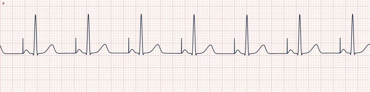
Example tracing, drawn by the lab's engine. Open the lab for a live, adjustable version.
At a glance
- Rate
- The programmed rate, perfectly regular
- Pacing spike
- One, immediately before each P wave
- P waves
- Follow every spike
- PR interval
- Normal, measured from the spike
Figures
Still images drawn by the lab's engine at 25 mm/s and 10 mm/mV, with the finding labeled. Select a figure to enlarge it.
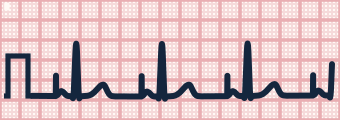
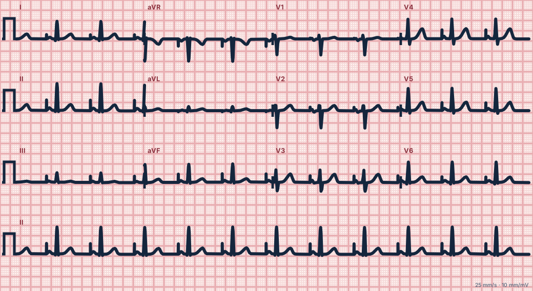
Scroll sideways to see the whole tracing, or select it to enlarge.
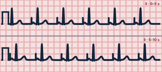
Scroll sideways to see the whole tracing, or select it to enlarge.
What the heart is doing
The pacemaker stimulates the right atrium at the programmed rate. The impulse spreads across the atria and through the patient's own AV node and His–Purkinje system.
Why it looks this way
This is an atrial paced rhythm because a pacing spike precedes each P wave, which then conducts normally to a narrow QRS.
Why it matters
Normal device function when each spike captures the atrium. Look for spikes without P waves or P waves without a QRS, which indicate failure to capture or new AV block.
What to look for
- A thin vertical spike right before each P wave.
- Every spike is followed by a P wave (atrial capture) and then a narrow QRS.
- The rhythm is metronome-regular at the device's set rate.
Criteria
- Rate
- The programmed rate, perfectly regular
- Pacing spike
- One, immediately before each P wave
- P waves
- Follow every spike
- PR interval
- Normal, measured from the spike
- QRS
- Narrow (< 120 ms)
Causes
- Sinus node dysfunction (sick sinus syndrome)
- Symptomatic sinus bradycardia
- Inability to raise heart rate with exertion
- Requires intact AV conduction
Can be mistaken for
- Sinus rhythm with artifact — the 'spikes' are not tied to the P waves
- AV sequential pacing — a second spike precedes a wide QRS Compare
- Ventricular pacing — the spike is followed directly by a wide QRS Compare
Advanced Down to the ion channel
What the cells are doing
Pacemaker, atrial and ventricular action potentials, in step with the tracing above, with sodium, calcium and potassium moving through their channels.
Frequently asked questions
What are the ECG criteria for atrial paced rhythm?
Rate: the programmed rate, perfectly regular. Pacing spike: one, immediately before each P wave. P waves: follow every spike. PR interval: normal, measured from the spike. QRS: narrow (< 120 ms).
How is atrial paced rhythm identified on an ECG?
This is an atrial paced rhythm because a pacing spike precedes each P wave, which then conducts normally to a narrow QRS.
What can atrial paced rhythm be mistaken for?
Sinus rhythm with artifact: the 'spikes' are not tied to the P waves. AV sequential pacing: a second spike precedes a wide QRS. Ventricular pacing: the spike is followed directly by a wide QRS.
What causes atrial paced rhythm?
Common causes: sinus node dysfunction (sick sinus syndrome); symptomatic sinus bradycardia; inability to raise heart rate with exertion; and requires intact AV conduction.
Sources
This page does not list page-specific sources yet.
Every source the site cites, with links