
Paced
Pacemaker failure to capture
The pacemaker fires, but some impulses fail to depolarize the heart. Spikes appear on time with no QRS after them.
Last updated
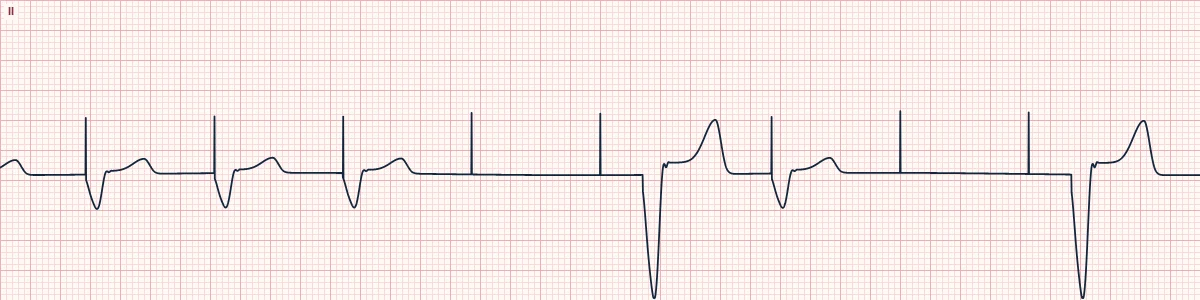
Example tracing, drawn by the lab's engine. Open the lab for a live, adjustable version.
At a glance
- Rate
- Slower than the programmed pacing rate
- Rhythm
- Irregular, with pauses
- Pacing spikes
- Regular; some not followed by a QRS
- QRS
- Wide after captured spikes; slow escape beats may appear
Figures
Still images drawn by the lab's engine at 25 mm/s and 10 mm/mV, with the finding labeled. Select a figure to enlarge it.
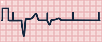
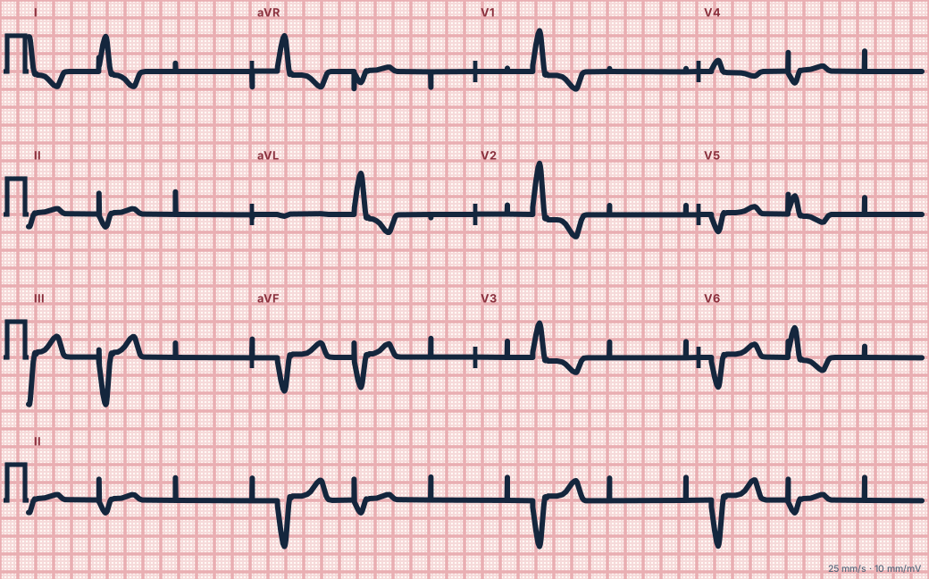
Scroll sideways to see the whole tracing, or select it to enlarge.
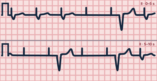
Scroll sideways to see the whole tracing, or select it to enlarge.
What the heart is doing
The stimulus is delivered but is too weak, or reaches tissue that cannot respond, so the ventricle does not depolarize. The heart then relies on any slow escape pacemaker it has.
Why it looks this way
This is failure to capture because pacing spikes occur on schedule but some are not followed by a QRS complex.
Why it matters
Potentially life-threatening in a patient who depends on the pacemaker. Treat the patient's bradycardia, be ready for external pacing, correct metabolic causes and arrange urgent device interrogation.
What to look for
- Spikes that stand alone with no wide QRS and T wave behind them.
- Spike-to-spike timing is regular, which shows the device is firing on schedule.
- Wide beats with no spike in front are the patient's own escape beats.
Criteria
- Rate
- Slower than the programmed pacing rate
- Rhythm
- Irregular, with pauses
- Pacing spikes
- Regular; some not followed by a QRS
- QRS
- Wide after captured spikes; slow escape beats may appear
- Pauses
- Lone spikes on a flat baseline
Causes
- Lead dislodgement or fracture
- Battery depletion
- Raised threshold from infarction or fibrosis at the lead tip
- Hyperkalemia, acidosis
- Antiarrhythmic drug effect
- Output set too low
Can be mistaken for
- Failure to pace (no output) — expected spikes are missing altogether
- Normal pacing — every spike is followed by a QRS
- Failure to sense — spikes appear where they should have been withheld Compare
Advanced Down to the ion channel
What the cells are doing
Pacemaker, atrial and ventricular action potentials, in step with the tracing above, with sodium, calcium and potassium moving through their channels.
Frequently asked questions
What are the ECG criteria for pacemaker failure to capture?
Rate: slower than the programmed pacing rate. Rhythm: irregular, with pauses. Pacing spikes: regular; some not followed by a QRS. QRS: wide after captured spikes; slow escape beats may appear. Pauses: lone spikes on a flat baseline.
How is pacemaker failure to capture identified on an ECG?
This is failure to capture because pacing spikes occur on schedule but some are not followed by a QRS complex.
What can pacemaker failure to capture be mistaken for?
Failure to pace (no output): expected spikes are missing altogether. Normal pacing: every spike is followed by a QRS. Failure to sense: spikes appear where they should have been withheld.
What causes pacemaker failure to capture?
Common causes: lead dislodgement or fracture; battery depletion; raised threshold from infarction or fibrosis at the lead tip; hyperkalemia, acidosis; antiarrhythmic drug effect; and output set too low.
Practise it in the ACLS Lab
Sources
This page does not list page-specific sources yet.
Every source the site cites, with links