
Bundle branch block
Nonspecific intraventricular conduction delay
The QRS is widened by slow conduction through the ventricular muscle, but the pattern fits neither right nor left bundle branch block.
Last updated
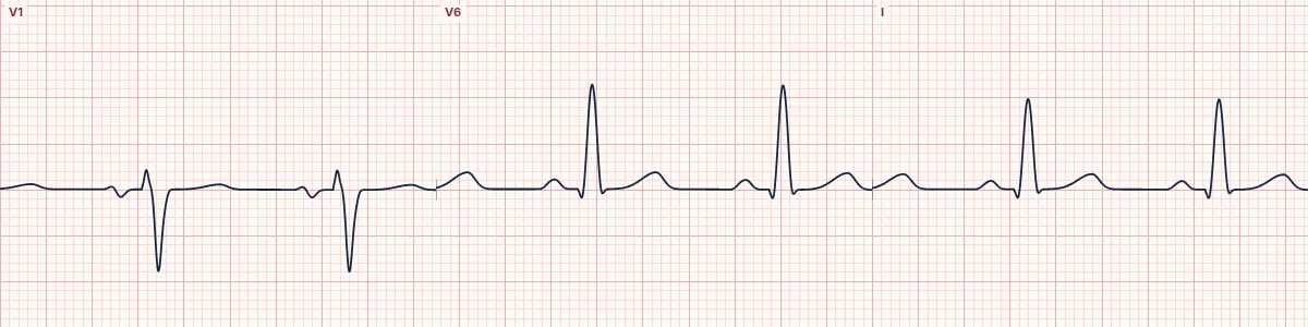
Example tracing, drawn by the lab's engine. Open the lab for a live, adjustable version.
At a glance
- QRS
- > 110 ms, often ≥ 120 ms
- V1
- No typical rsR′ or broad QS
- V6
- No typical wide S or broad notched R
- ST–T
- Flattened or nonspecific changes
Figures
Still images drawn by the lab's engine at 25 mm/s and 10 mm/mV, with the finding labeled. Select a figure to enlarge it.
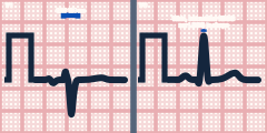
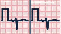
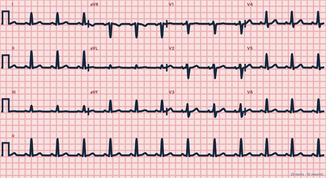
Scroll sideways to see the whole tracing, or select it to enlarge.
What the heart is doing
The bundle branches conduct, but the impulse spreads slowly through the Purkinje network and muscle, because of scarring, hypertrophy or depressed sodium channels. The whole QRS is uniformly prolonged.
Why it looks this way
This is a nonspecific intraventricular conduction delay because the QRS is wide without the V1 and V6 features of right or left bundle branch block.
Why it matters
Chronic IVCD marks diseased ventricular muscle. Newly widened QRS complexes should prompt an immediate search for hyperkalemia or drug toxicity, both of which are reversible and dangerous.
What to look for
- A wide QRS that keeps a roughly normal shape, as if stretched sideways.
- Check V1 and V6: neither shows the classic RBBB or LBBB pattern.
- If it is new, look for peaked T waves (hyperkalemia) or a tall R in aVR (sodium-channel blockade).
Criteria
- QRS
- > 110 ms, often ≥ 120 ms
- V1
- No typical rsR′ or broad QS
- V6
- No typical wide S or broad notched R
- ST–T
- Flattened or nonspecific changes
Causes
- Cardiomyopathy and myocardial scar
- Left ventricular hypertrophy
- Hyperkalemia
- Sodium-channel blocking drugs
- Severe hypothermia
Can be mistaken for
Advanced Down to the ion channel
What the cells are doing
Pacemaker, atrial and ventricular action potentials, in step with the tracing above, with sodium, calcium and potassium moving through their channels.
Frequently asked questions
What are the ECG criteria for nonspecific intraventricular conduction delay?
QRS: > 110 ms, often ≥ 120 ms. V1: no typical rsR′ or broad QS. V6: no typical wide S or broad notched R. ST–T: flattened or nonspecific changes.
How is nonspecific intraventricular conduction delay identified on an ECG?
This is a nonspecific intraventricular conduction delay because the QRS is wide without the V1 and V6 features of right or left bundle branch block.
What can nonspecific intraventricular conduction delay be mistaken for?
LBBB: broad notched R in V6 and QS in V1. RBBB: rsR′ in V1 and wide S in V6. WPW: short PR with a slurred delta wave.
What causes nonspecific intraventricular conduction delay?
Common causes: cardiomyopathy and myocardial scar; left ventricular hypertrophy; hyperkalemia; sodium-channel blocking drugs; and severe hypothermia.
Sources
This page does not list page-specific sources yet.
Every source the site cites, with links