
Bundle branch block
Incomplete right bundle branch block
Slight delay in the right bundle branch produces the RBBB shape in V1 while the QRS stays under 120 ms. Commonly a normal variant.
Last updated
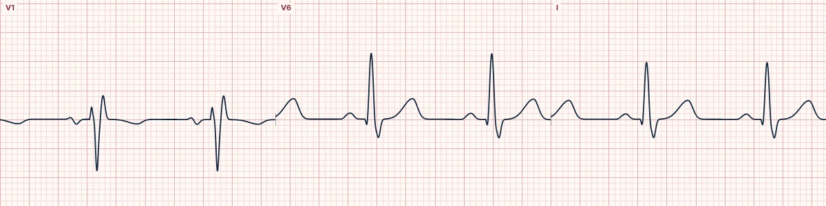
Example tracing, drawn by the lab's engine. Open the lab for a live, adjustable version.
At a glance
- QRS
- 110–119 ms in adults; 100–110 ms in children
- V1
- rsr′ or rsR′ pattern
- V6 and I
- Small terminal S wave
- ST–T
- Usually normal
Figures
Still images drawn by the lab's engine at 25 mm/s and 10 mm/mV, with the finding labeled. Select a figure to enlarge it.
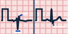
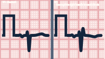
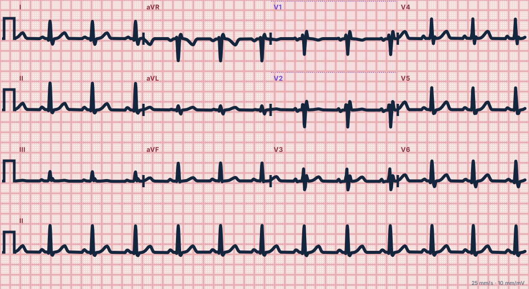
Scroll sideways to see the whole tracing, or select it to enlarge.
What the heart is doing
Conduction down the right bundle is slowed but not blocked, so the last part of the right ventricle depolarizes slightly late. This adds a small terminal force toward V1.
Why it looks this way
This is incomplete RBBB because V1 shows an rsr′ pattern while the QRS is between 110 and 119 ms.
Why it matters
Usually harmless and needs no action. In a breathless patient or one with a murmur, consider right heart strain or an atrial septal defect.
What to look for
- A small second upward deflection (r′) at the end of the QRS in V1.
- Measure the QRS: under 120 ms, so it does not meet criteria for complete RBBB.
- Check that the ST segment in V1–V2 is not elevated, which would suggest Brugada pattern.
Criteria
- QRS
- 110–119 ms in adults; 100–110 ms in children
- V1
- rsr′ or rsR′ pattern
- V6 and I
- Small terminal S wave
- ST–T
- Usually normal
Causes
- Normal variant in the young and athletic
- Atrial septal defect
- Right ventricular volume or pressure load
- V1 electrode placed too high
- Early conduction disease
Can be mistaken for
- Complete RBBB — QRS 120 ms or more with a broad S in V6 Compare
- Brugada pattern, type 2 — saddle-shaped ST elevation in V1–V2 Compare
- High V1 placement — r′ with an inverted P wave in V1
Advanced Down to the ion channel
What the cells are doing
Pacemaker, atrial and ventricular action potentials, in step with the tracing above, with sodium, calcium and potassium moving through their channels.
Frequently asked questions
What are the ECG criteria for incomplete right bundle branch block?
QRS: 110–119 ms in adults; 100–110 ms in children. V1: rsr′ or rsR′ pattern. V6 and I: small terminal S wave. ST–T: usually normal.
How is incomplete right bundle branch block identified on an ECG?
This is incomplete RBBB because V1 shows an rsr′ pattern while the QRS is between 110 and 119 ms.
What can incomplete right bundle branch block be mistaken for?
Complete RBBB: QRS 120 ms or more with a broad S in V6. Brugada pattern, type 2: saddle-shaped ST elevation in V1–V2. High V1 placement: r′ with an inverted P wave in V1.
What causes incomplete right bundle branch block?
Common causes: normal variant in the young and athletic; atrial septal defect; right ventricular volume or pressure load; V1 electrode placed too high; and early conduction disease.
Sources
This page does not list page-specific sources yet.
Every source the site cites, with links