
Bundle branch block
Left bundle branch block
The left bundle branch does not conduct, so the left ventricle is activated late and slowly from the right side. The QRS is wide, with ST–T changes opposite to it.
Last updated
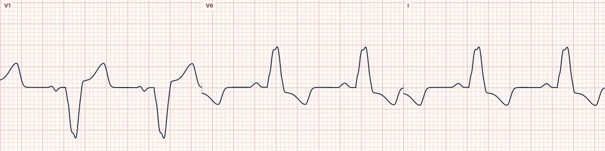
Example tracing, drawn by the lab's engine. Open the lab for a live, adjustable version.
At a glance
- QRS
- ≥ 120 ms
- V1
- Deep, broad QS or rS
- V6, I, aVL
- Broad, notched R wave with no septal q
- ST–T
- Opposite to the QRS and in proportion: ST elevation in V1–V3 well under a quarter of the S-wave depth, T inversion laterally
Figures
Still images drawn by the lab's engine at 25 mm/s and 10 mm/mV, with the finding labeled. Select a figure to enlarge it.
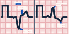
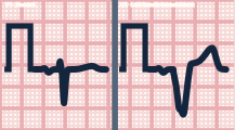
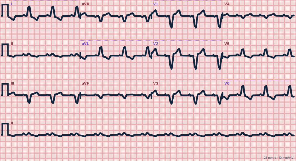
Scroll sideways to see the whole tracing, or select it to enlarge.
What the heart is doing
The septum depolarizes right to left, the reverse of normal, removing the septal q wave. The impulse then spreads slowly through muscle to the left ventricle, giving a broad leftward, posterior force.
Why it looks this way
This is LBBB because the QRS is at least 120 ms with a broad notched R in I and V6, a deep QS in V1 and no septal q waves.
Why it matters
Almost always signals structural heart disease. It hides the usual signs of infarction. Use the Sgarbossa criteria as modified by Dr. Stephen W. Smith: concordant ST elevation, V1–V3 ST depression of 1 mm or more, or ST elevation at least 25% of the S-wave depth (ST/S ratio −0.25 or lower).
What to look for
- V1 is almost entirely negative and wide; V6 is entirely positive, broad and notched.
- The small septal q wave normally seen in I and V6 is gone.
- ST segments should point away from the QRS; ST deviation in the same direction is abnormal.
Criteria
- QRS
- ≥ 120 ms
- V1
- Deep, broad QS or rS
- V6, I, aVL
- Broad, notched R wave with no septal q
- ST–T
- Opposite to the QRS and in proportion: ST elevation in V1–V3 well under a quarter of the S-wave depth, T inversion laterally
- Axis
- Normal or leftward
Causes
- Hypertension and left ventricular hypertrophy
- Coronary artery disease, infarction
- Dilated cardiomyopathy
- Aortic valve disease
- Degenerative conduction disease
Can be mistaken for
- Ventricular paced rhythm — pacing spikes before each QRS Compare
- Left ventricular hypertrophy — QRS under 120 ms with septal q waves preserved Compare
- WPW type B — short PR and delta wave Compare
Advanced Down to the ion channel
What the cells are doing
Pacemaker, atrial and ventricular action potentials, in step with the tracing above, with sodium, calcium and potassium moving through their channels.
Frequently asked questions
What are the ECG criteria for left bundle branch block?
QRS: ≥ 120 ms. V1: deep, broad QS or rS. V6, I, aVL: broad, notched R wave with no septal q. ST–T: opposite to the QRS and in proportion: ST elevation in V1–V3 well under a quarter of the S-wave depth, T inversion laterally. Axis: normal or leftward.
How is left bundle branch block identified on an ECG?
This is LBBB because the QRS is at least 120 ms with a broad notched R in I and V6, a deep QS in V1 and no septal q waves.
What can left bundle branch block be mistaken for?
Ventricular paced rhythm: pacing spikes before each QRS. Left ventricular hypertrophy: QRS under 120 ms with septal q waves preserved. WPW type B: short PR and delta wave.
What causes left bundle branch block?
Common causes: hypertension and left ventricular hypertrophy; coronary artery disease, infarction; dilated cardiomyopathy; aortic valve disease; and degenerative conduction disease.
Sources
This page does not list page-specific sources yet.
Every source the site cites, with links