
Coronary occlusion (STEMI)
Left circumflex occlusion
Inferolateral and posterior STEMI
Blockage of the left circumflex artery (LCx), which runs around the back of the heart. It infarcts the lateral, inferior and posterior walls of the left ventricle.
Last updated
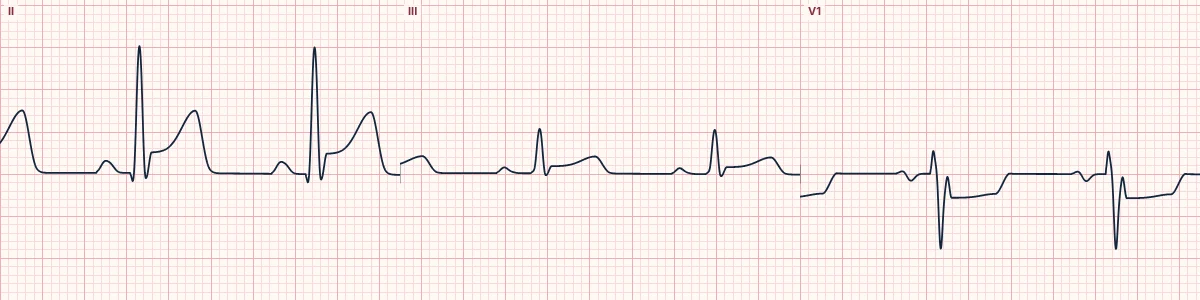
Example tracing, drawn by the lab's engine. Open the lab for a live, adjustable version.
At a glance
- Artery
- Left circumflex
- ST elevation
- II, III, aVF, V5, V6, V7, V8, V9
- Reciprocal ST depression
- V1, V2, V3
- Pattern
- ST elevation in II at least equal to III
Figures
Still images drawn by the lab's engine at 25 mm/s and 10 mm/mV, with the finding labeled. Select a figure to enlarge it.
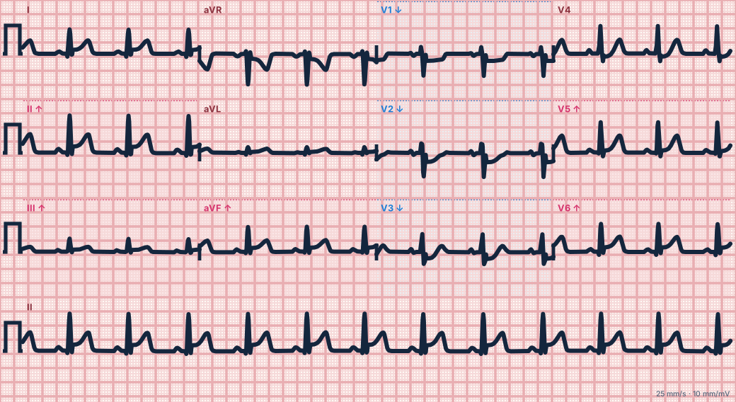
Scroll sideways to see the whole tracing, or select it to enlarge.
What the heart is doing
Injury to the lateral, inferior and posterior walls produces an ST vector pointing down, left and backward. Inferior, lateral and posterior leads face it; V1–V3 see it in mirror image.
Why it looks this way
This is a circumflex occlusion because inferior and lateral (V5–V6) ST elevation is paired with ST depression in V1–V3 and elevation in V7–V9.
Why it matters
The circumflex territory is poorly seen by the standard 12 leads, so the ECG understates the infarct. Posterior leads help. Treat as a STEMI needing immediate reperfusion; watch for mitral regurgitation.
What to look for
- Inferior ST elevation that is no greater in III than in II, with V5–V6 also raised.
- ST depression in V1–V3 signals posterior wall injury; add leads V7–V9.
- Lead I is flat or slightly elevated, not depressed as in RCA occlusion.
Criteria
- Artery
- Left circumflex
- ST elevation
- II, III, aVF, V5, V6, V7, V8, V9
- Reciprocal ST depression
- V1, V2, V3
- Pattern
- ST elevation in II at least equal to III
- V1–V2
- Tall R wave and upright T may develop
Causes
- Plaque rupture with thrombosis
- Coronary artery spasm or stimulant use
- Spontaneous coronary dissection
- Coronary embolism
Can be mistaken for
- RCA occlusion — elevation greater in III than II with depression in I and aVL
- Anterior ischemia — V1–V3 depression without inferior or posterior elevation
- Pericarditis — diffuse elevation with PR depression and no V1–V3 depression Compare
Advanced Down to the ion channel
What the cells are doing
Pacemaker, atrial and ventricular action potentials, in step with the tracing above, with sodium, calcium and potassium moving through their channels.
Frequently asked questions
What are the ECG criteria for left circumflex occlusion?
Artery: left circumflex. ST elevation: II, III, aVF, V5, V6, V7, V8, V9. Reciprocal ST depression: V1, V2, V3. Pattern: ST elevation in II at least equal to III. V1–V2: tall R wave and upright T may develop.
How is left circumflex occlusion identified on an ECG?
This is a circumflex occlusion because inferior and lateral (V5–V6) ST elevation is paired with ST depression in V1–V3 and elevation in V7–V9.
What can left circumflex occlusion be mistaken for?
RCA occlusion: elevation greater in III than II with depression in I and aVL. Anterior ischemia: V1–V3 depression without inferior or posterior elevation. Pericarditis: diffuse elevation with PR depression and no V1–V3 depression.
What causes left circumflex occlusion?
Common causes: plaque rupture with thrombosis; coronary artery spasm or stimulant use; spontaneous coronary dissection; and coronary embolism.
Sources
This page does not list page-specific sources yet.
Every source the site cites, with links