
Coronary occlusion (STEMI)
First diagonal (D1) occlusion
High lateral STEMI
Blockage of the first diagonal branch (D1) of the LAD, which supplies the high lateral wall of the left ventricle. The changes are small and easily missed.
Last updated
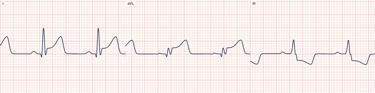
Example tracing, drawn by the lab's engine. Open the lab for a live, adjustable version.
At a glance
- Artery
- First diagonal branch of the LAD
- ST elevation
- I, aVL, V2
- Reciprocal ST depression
- III, aVF
- T waves
- Upright and enlarged in I and aVL; inverted in III
Figures
Still images drawn by the lab's engine at 25 mm/s and 10 mm/mV, with the finding labeled. Select a figure to enlarge it.
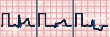
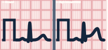
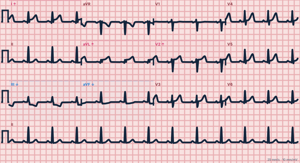
Scroll sideways to see the whole tracing, or select it to enlarge.
What the heart is doing
Injury to the high lateral wall produces an ST vector pointing up and to the left, toward aVL and I. Lead III faces almost exactly the opposite way and shows the mirror-image depression.
Why it looks this way
This is a first diagonal occlusion because ST elevation in I, aVL and V2 is matched by reciprocal ST depression in III and aVF.
Why it matters
A true STEMI that often fails to meet size criteria because aVL has low voltage. Recognize the pattern, obtain serial ECGs and treat as an acute coronary occlusion needing urgent reperfusion.
What to look for
- Start with aVL: any ST elevation there with ST depression in III is an occlusion until proven otherwise.
- Chest-lead changes are modest; the diagnosis rests on I, aVL and the mirror-image depression in III.
- Judge the ST elevation against the small QRS in aVL, not against an absolute number.
Criteria
- Artery
- First diagonal branch of the LAD
- ST elevation
- I, aVL, V2
- Reciprocal ST depression
- III, aVF
- T waves
- Upright and enlarged in I and aVL; inverted in III
- Amplitude
- Often only 1 mm of elevation in aVL
Causes
- Plaque rupture with thrombosis
- Coronary artery spasm or stimulant use
- Spontaneous coronary dissection
- Coronary embolism
Can be mistaken for
- Left ventricular hypertrophy — strain depression, not elevation, in aVL Compare
- Proximal LAD occlusion — ST elevation extends across V1–V6 Compare
- Arm lead reversal — P wave inverted in lead I Compare
Advanced Down to the ion channel
What the cells are doing
Pacemaker, atrial and ventricular action potentials, in step with the tracing above, with sodium, calcium and potassium moving through their channels.
Frequently asked questions
What are the ECG criteria for first diagonal (D1) occlusion?
Artery: first diagonal branch of the LAD. ST elevation: I, aVL, V2. Reciprocal ST depression: III, aVF. T waves: upright and enlarged in I and aVL; inverted in III. Amplitude: often only 1 mm of elevation in aVL.
How is first diagonal (D1) occlusion identified on an ECG?
This is a first diagonal occlusion because ST elevation in I, aVL and V2 is matched by reciprocal ST depression in III and aVF.
What can first diagonal (D1) occlusion be mistaken for?
Left ventricular hypertrophy: strain depression, not elevation, in aVL. Proximal LAD occlusion: ST elevation extends across V1–V6. Arm lead reversal: P wave inverted in lead I.
What causes first diagonal (D1) occlusion?
Common causes: plaque rupture with thrombosis; coronary artery spasm or stimulant use; spontaneous coronary dissection; and coronary embolism.
Sources
This page does not list page-specific sources yet.
Every source the site cites, with links