
Coronary occlusion (STEMI)
Posterior branch occlusion
Isolated posterior MI
Blockage of a posterior branch, usually of the left circumflex artery (LCx), infarcting the posterior wall. No standard lead faces it, so it shows only as mirror-image changes in V1–V3.
Last updated
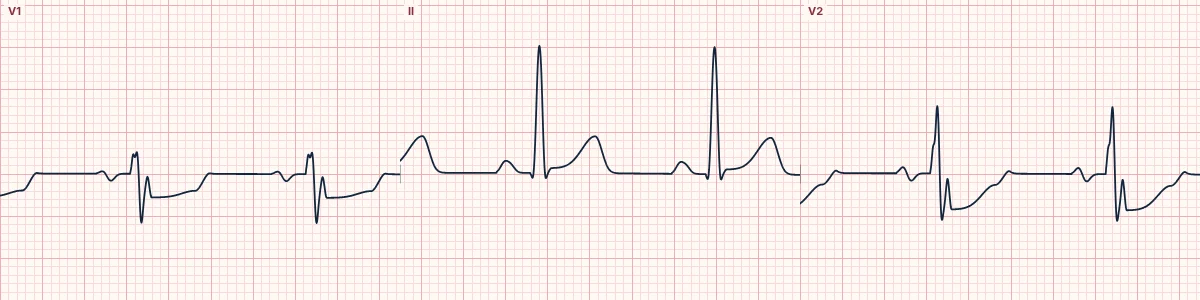
Example tracing, drawn by the lab's engine. Open the lab for a live, adjustable version.
At a glance
- Artery
- Distal circumflex (posterior branch)
- ST elevation
- V7, V8, V9
- Reciprocal ST depression
- V1, V2, V3
- V1–V3
- Horizontal ST depression with upright (or terminally upright) T waves; a tall, broad R as it evolves
Figures
Still images drawn by the lab's engine at 25 mm/s and 10 mm/mV, with the finding labeled. Select a figure to enlarge it.
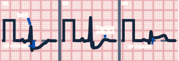
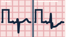
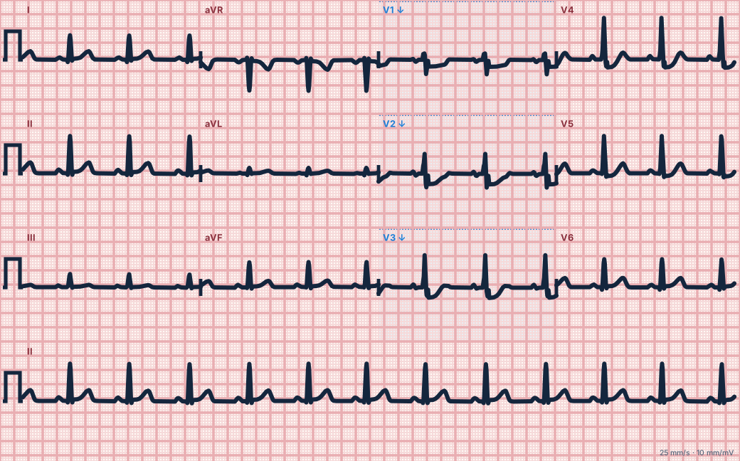
Scroll sideways to see the whole tracing, or select it to enlarge.
What the heart is doing
Posterior wall injury produces an ST vector pointing straight backward, away from the chest leads. V1–V3 record it upside down: ST depression for elevation and tall R waves for Q waves.
Why it looks this way
This is a posterior occlusion because ST depression with tall R waves in V1–V3 mirrors ST elevation recorded in the posterior leads V7–V9.
Why it matters
Frequently mislabeled as non-ST-elevation ischemia, delaying reperfusion. ST depression maximal in V1–V3 with symptoms should be treated as a STEMI equivalent and confirmed with posterior leads.
What to look for
- Horizontal ST depression greatest in V1–V3 should always prompt posterior leads.
- A tall, broad R wave in V2 (R larger than S) is a posterior Q wave seen from the front.
- Place V7–V9 on the back at the level of V6; small elevations count.
Criteria
- Artery
- Distal circumflex (posterior branch)
- ST elevation
- V7, V8, V9
- Reciprocal ST depression
- V1, V2, V3
- V1–V3
- Horizontal ST depression with upright (or terminally upright) T waves; a tall, broad R as it evolves
- Threshold
- ≥ 0.5 mm of elevation in V7–V9 is significant
Causes
- Plaque rupture with thrombosis
- Coronary artery spasm or stimulant use
- Spontaneous coronary dissection
- Coronary embolism
Can be mistaken for
- Anterior subendocardial ischemia — depression usually maximal in V4–V6
- RBBB — rsR′ in V1 with a wide QRS Compare
- Right ventricular hypertrophy — tall R in V1 with right axis deviation Compare
Advanced Down to the ion channel
What the cells are doing
Pacemaker, atrial and ventricular action potentials, in step with the tracing above, with sodium, calcium and potassium moving through their channels.
Frequently asked questions
What are the ECG criteria for posterior branch occlusion?
Artery: distal circumflex (posterior branch). ST elevation: V7, V8, V9. Reciprocal ST depression: V1, V2, V3. V1–V3: horizontal ST depression with upright (or terminally upright) T waves; a tall, broad R as it evolves. Threshold: ≥ 0.5 mm of elevation in V7–V9 is significant.
How is posterior branch occlusion identified on an ECG?
This is a posterior occlusion because ST depression with tall R waves in V1–V3 mirrors ST elevation recorded in the posterior leads V7–V9.
What can posterior branch occlusion be mistaken for?
Anterior subendocardial ischemia: depression usually maximal in V4–V6. RBBB: rsR′ in V1 with a wide QRS. Right ventricular hypertrophy: tall R in V1 with right axis deviation.
What causes posterior branch occlusion?
Common causes: plaque rupture with thrombosis; coronary artery spasm or stimulant use; spontaneous coronary dissection; and coronary embolism.
Sources
This page does not list page-specific sources yet.
Every source the site cites, with links