
Leads and axis
Finding the frontal QRS axis
Leads I and aVF
The axis is the average direction of ventricular activation in the frontal plane. Normal is −30° to +90°. Leads I and aVF give the quadrant in seconds; lead II settles the borderline.
Last updated
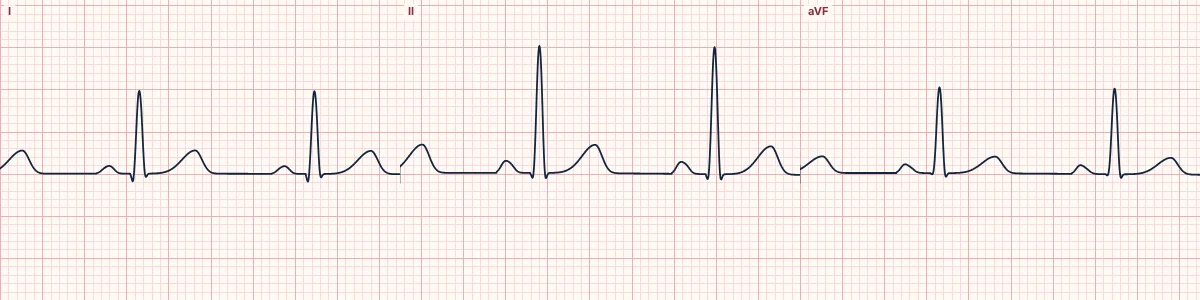
Example tracing, drawn by the lab's engine. Open the lab for a live, adjustable version.
At a glance
- Normal
- −30° to +90°
- Left axis deviation
- −30° to −90°
- Right axis deviation
- +90° to +180°
- Extreme axis
- −90° to ±180°
Figures
Still images drawn by the lab's engine at 25 mm/s and 10 mm/mV, with the finding labeled. Select a figure to enlarge it.
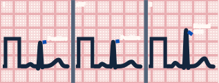
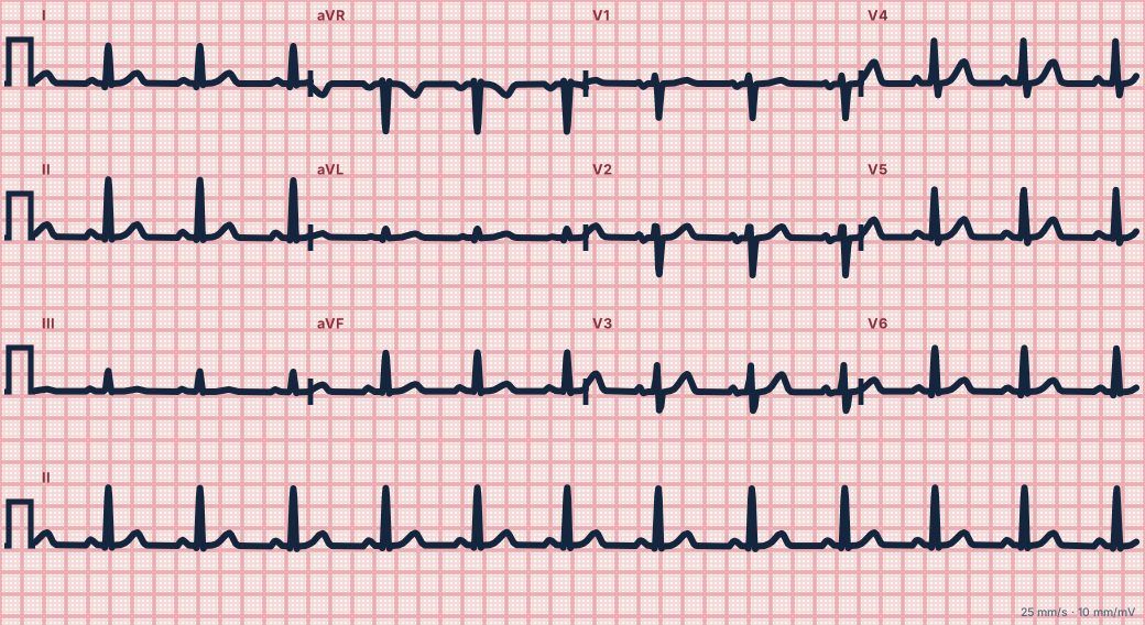
Scroll sideways to see the whole tracing, or select it to enlarge.
Quadrant method
If the QRS is mainly positive in both I and aVF, the axis is normal (0° to +90°). Positive in I and negative in aVF means the axis is leftward; then look at lead II. If II is mainly positive, the axis is still normal (0° to −30°); if II is negative, there is left axis deviation.
Isoelectric lead method
Find the limb lead whose QRS is most nearly equal up and down. The axis is at right angles to it, toward whichever of the two perpendicular leads is positive. This gives the axis to within about 15°.
Try it
In the lab, pause the tracing and drag the axis arrow on the hexaxial wheel. Every limb lead redraws as you move it, which is the quickest way to feel how the leads change with the axis.
How to read it
- Look at I and aVF first.
- If I is positive and aVF negative, check lead II.
- Find the most isoelectric limb lead to refine the angle.
Pitfalls
- Swapped arm electrodes produce a false right axis.
- Right bundle branch block's late forces are excluded when quoting the axis.
Key numbers
- Normal
- −30° to +90°
- Left axis deviation
- −30° to −90°
- Right axis deviation
- +90° to +180°
- Extreme axis
- −90° to ±180°
Advanced Down to the ion channel
What the cells are doing
Pacemaker, atrial and ventricular action potentials, in step with the tracing above, with sodium, calcium and potassium moving through their channels.
Frequently asked questions
Finding the frontal QRS axis: what is the key idea?
The axis is the average direction of ventricular activation in the frontal plane. Normal is −30° to +90°. Leads I and aVF give the quadrant in seconds; lead II settles the borderline.
Finding the frontal QRS axis: what are the key numbers?
Normal: −30° to +90°; Left axis deviation: −30° to −90°; Right axis deviation: +90° to +180°; Extreme axis: −90° to ±180°.
Finding the frontal QRS axis: how do you apply it to an ECG?
Look at I and aVF first. If I is positive and aVF negative, check lead II. Find the most isoelectric limb lead to refine the angle.
Finding the frontal QRS axis: what are the common pitfalls?
Swapped arm electrodes produce a false right axis. Right bundle branch block's late forces are excluded when quoting the axis.
Sources
This page does not list page-specific sources yet.
Every source the site cites, with links