
Leads and axis
Axis deviation and its causes
Left, right and extreme
A shifted axis is a clue rather than a diagnosis: it points to blocked fascicles, enlarged ventricles, old infarcts, ventricular origin or misplaced electrodes.
Last updated
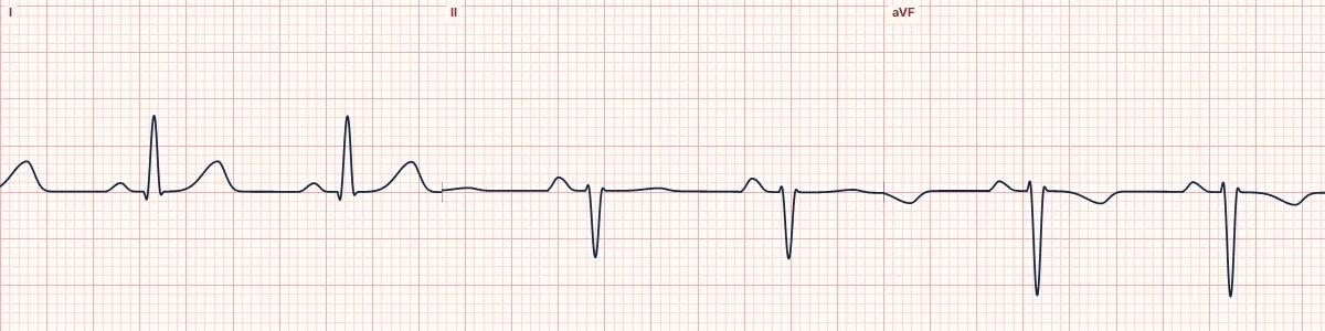
Example tracing, drawn by the lab's engine. Open the lab for a live, adjustable version.
At a glance
- LAD
- I positive, aVF negative, II negative
- RAD
- I negative, aVF positive
- Extreme
- I negative, aVF negative
Figures
Still images drawn by the lab's engine at 25 mm/s and 10 mm/mV, with the finding labeled. Select a figure to enlarge it.
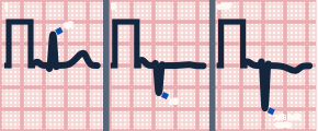
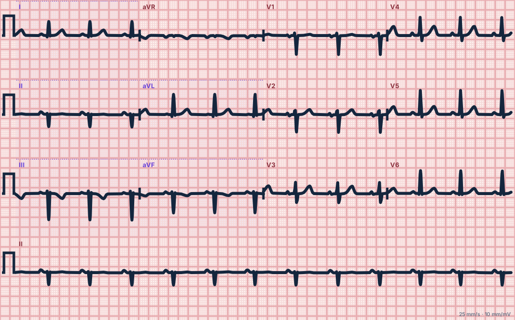
Scroll sideways to see the whole tracing, or select it to enlarge.
Left axis deviation
Commonly left anterior fascicular block, which gives qR in I and aVL and rS in II, III and aVF. Also inferior myocardial infarction, left ventricular hypertrophy, left bundle branch block, pre-excitation, ventricular rhythms and paced rhythms from the right ventricular apex.
Right axis deviation
Normal in children and tall thin adults. Pathologically, right ventricular hypertrophy, acute or chronic pulmonary disease including pulmonary embolism, lateral myocardial infarction, left posterior fascicular block (a diagnosis of exclusion), dextrocardia and swapped arm leads.
Extreme axis
An axis between −90° and ±180° (northwest) means activation is travelling from the apex up toward the base. In a wide-complex tachycardia it strongly favours ventricular tachycardia.
How to read it
- Name the axis, then ask which of the causes fits the rest of the ECG.
- Compare with old tracings: a new axis shift is more important than a stable one.
Pitfalls
- Left posterior fascicular block should only be called when right ventricular and lateral causes are excluded.
Key numbers
- LAD
- I positive, aVF negative, II negative
- RAD
- I negative, aVF positive
- Extreme
- I negative, aVF negative
Advanced Down to the ion channel
What the cells are doing
Pacemaker, atrial and ventricular action potentials, in step with the tracing above, with sodium, calcium and potassium moving through their channels.
Frequently asked questions
Axis deviation and its causes: what is the key idea?
A shifted axis is a clue rather than a diagnosis: it points to blocked fascicles, enlarged ventricles, old infarcts, ventricular origin or misplaced electrodes.
Axis deviation and its causes: what are the key numbers?
LAD: I positive, aVF negative, II negative; RAD: I negative, aVF positive; Extreme: I negative, aVF negative.
Axis deviation and its causes: how do you apply it to an ECG?
Name the axis, then ask which of the causes fits the rest of the ECG. Compare with old tracings: a new axis shift is more important than a stable one.
Axis deviation and its causes: what are the common pitfalls?
Left posterior fascicular block should only be called when right ventricular and lateral causes are excluded.
Sources
This page does not list page-specific sources yet.
Every source the site cites, with links