
Rate-related aberrancy
Rate-related LBBB aberrancy
The left bundle fails only when the heart rate rises above a critical level or a beat arrives early, so LBBB comes and goes with the rate.
Last updated
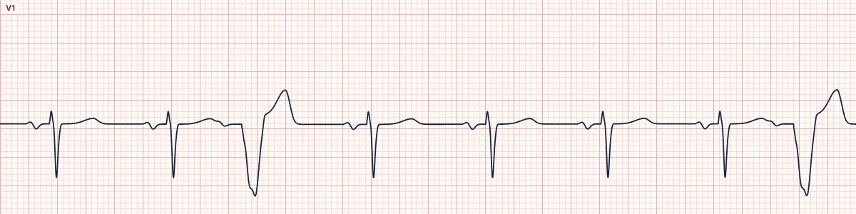
Example tracing, drawn by the lab's engine. Open the lab for a live, adjustable version.
At a glance
- QRS
- Wide (≥ 120 ms) on fast or early beats only
- V1
- Broad QS or rS on the aberrant beats
- V6 and I
- Broad notched R on the aberrant beats
- Timing
- Appears at short R–R; narrows as the rate slows
Figures
Still images drawn by the lab's engine at 25 mm/s and 10 mm/mV, with the finding labeled. Select a figure to enlarge it.
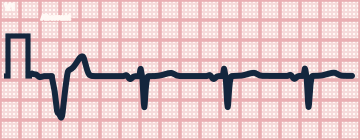
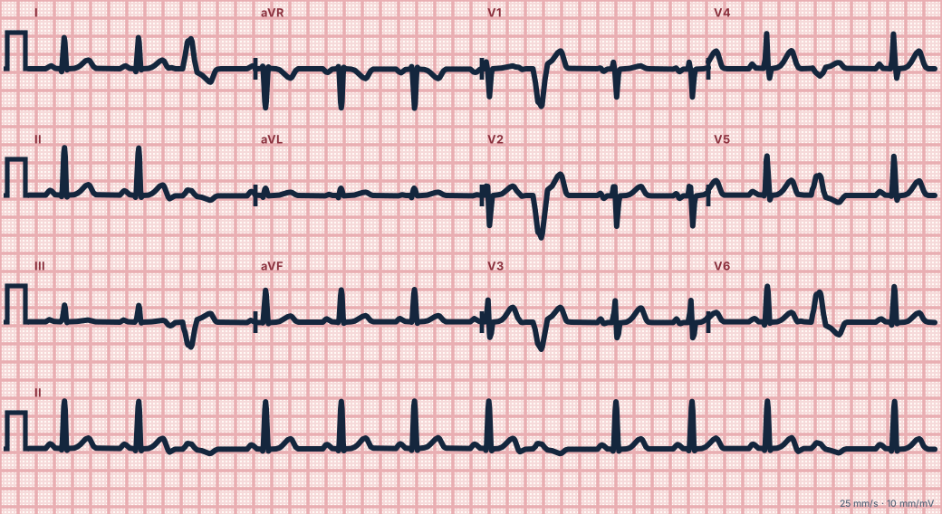
Scroll sideways to see the whole tracing, or select it to enlarge.
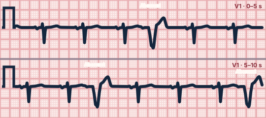
Scroll sideways to see the whole tracing, or select it to enlarge.
What the heart is doing
A diseased left bundle needs longer to recover than the right. When the cycle shortens below that recovery time, the impulse travels down the right bundle only and reaches the left ventricle late.
Why it looks this way
This is rate-related LBBB aberrancy because an LBBB pattern appears on beats with a short R–R interval and resolves when the rate slows.
Why it matters
More often linked to heart disease than RBBB aberrancy. A tachycardia with rate-related LBBB can look like VT, and its ST–T changes can mimic ischemia.
What to look for
- Compare the QRS at short and long R–R intervals: wide when fast, narrow when slow.
- Each wide beat still has the same P wave and PR relationship as the narrow ones.
- The wide beats have a typical LBBB shape, not a bizarre ventricular one.
Criteria
- QRS
- Wide (≥ 120 ms) on fast or early beats only
- V1
- Broad QS or rS on the aberrant beats
- V6 and I
- Broad notched R on the aberrant beats
- Timing
- Appears at short R–R; narrows as the rate slows
Causes
- Underlying left bundle disease
- Coronary artery disease
- Hypertension
- Cardiomyopathy
- Tachycardia from any cause
Can be mistaken for
- Ventricular tachycardia or PVCs — no preceding P wave, and AV dissociation
- Fixed LBBB — wide at every rate Compare
- Intermittent WPW type B — short PR and delta wave on the wide beats
Advanced Down to the ion channel
What the cells are doing
Pacemaker, atrial and ventricular action potentials, in step with the tracing above, with sodium, calcium and potassium moving through their channels.
Frequently asked questions
What are the ECG criteria for rate-related LBBB aberrancy?
QRS: wide (≥ 120 ms) on fast or early beats only. V1: broad QS or rS on the aberrant beats. V6 and I: broad notched R on the aberrant beats. Timing: appears at short R–R; narrows as the rate slows.
How is rate-related LBBB aberrancy identified on an ECG?
This is rate-related LBBB aberrancy because an LBBB pattern appears on beats with a short R–R interval and resolves when the rate slows.
What can rate-related LBBB aberrancy be mistaken for?
Ventricular tachycardia or PVCs: no preceding P wave, and AV dissociation. Fixed LBBB: wide at every rate. Intermittent WPW type B: short PR and delta wave on the wide beats.
What causes rate-related LBBB aberrancy?
Common causes: underlying left bundle disease; coronary artery disease; hypertension; cardiomyopathy; and tachycardia from any cause.
Sources
This page does not list page-specific sources yet.
Every source the site cites, with links