
Ischemia and infarction
Reciprocal changes
The mirror image
ST elevation over an injured wall appears as ST depression in leads looking from the opposite side. Its presence makes acute occlusion far more likely and helps separate it from pericarditis and early repolarization.
Last updated
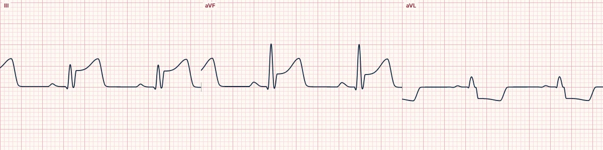
Example tracing, drawn by the lab's engine. Open the lab for a live, adjustable version.
At a glance
- Inferior STE
- Depression in aVL (and I)
- High lateral STE
- Depression in III and aVF
- Posterior
- Depression in V1–V3
- Pericarditis
- No reciprocal depression except aVR, V1
Figures
Still images drawn by the lab's engine at 25 mm/s and 10 mm/mV, with the finding labeled. Select a figure to enlarge it.
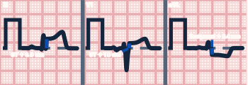
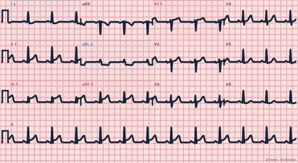
Scroll sideways to see the whole tracing, or select it to enlarge.
Why it happens
The current of injury points toward the injured wall. Leads facing that wall record elevation; leads facing away record the same shift upside down.
Classic pairs
Inferior elevation (II, III, aVF) with depression in aVL, often the earliest sign of inferior occlusion. High lateral elevation (I, aVL) with inferior depression. Posterior infarction shows only the reciprocal view: horizontal ST depression in V1–V3.
Using it
Diffuse ST elevation without reciprocal depression (except in aVR and V1) favours pericarditis. Subtle inferior elevation with clear depression in aVL favours occlusion.
How to read it
- For every lead with elevation, look at the leads opposite.
- Treat ST depression in aVL with inferior elevation as reciprocal until proven otherwise.
Pitfalls
- Widespread ST depression with elevation in aVR is a separate pattern of diffuse subendocardial ischemia, not a reciprocal change.
Key numbers
- Inferior STE
- Depression in aVL (and I)
- High lateral STE
- Depression in III and aVF
- Posterior
- Depression in V1–V3
- Pericarditis
- No reciprocal depression except aVR, V1
Advanced Down to the ion channel
What the cells are doing
Pacemaker, atrial and ventricular action potentials, in step with the tracing above, with sodium, calcium and potassium moving through their channels.
Frequently asked questions
Reciprocal changes: what is the key idea?
ST elevation over an injured wall appears as ST depression in leads looking from the opposite side. Its presence makes acute occlusion far more likely and helps separate it from pericarditis and early repolarization.
Reciprocal changes: what are the key numbers?
Inferior STE: Depression in aVL (and I); High lateral STE: Depression in III and aVF; Posterior: Depression in V1–V3; Pericarditis: No reciprocal depression except aVR, V1.
Reciprocal changes: how do you apply it to an ECG?
For every lead with elevation, look at the leads opposite. Treat ST depression in aVL with inferior elevation as reciprocal until proven otherwise.
Reciprocal changes: what are the common pitfalls?
Widespread ST depression with elevation in aVR is a separate pattern of diffuse subendocardial ischemia, not a reciprocal change.
Sources
This page does not list page-specific sources yet.
Every source the site cites, with links