
Ischemia and infarction
Occlusion in left bundle branch block
Sgarbossa and Smith criteria
Left bundle branch block and ventricular pacing distort the ST segment by themselves, hiding acute occlusion. The Sgarbossa criteria, as modified by Smith, look for ST shifts that the block cannot explain.
Last updated
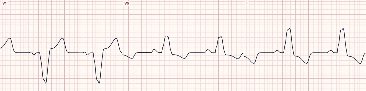
Example tracing, drawn by the lab's engine. Open the lab for a live, adjustable version.
At a glance
- Concordant STE
- ≥ 1 mm, any lead
- Concordant STD
- ≥ 1 mm in V1–V3
- Excessive discordance
- ST/S ratio ≤ −0.25
Figures
Still images drawn by the lab's engine at 25 mm/s and 10 mm/mV, with the finding labeled. Select a figure to enlarge it.
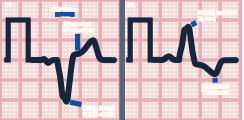
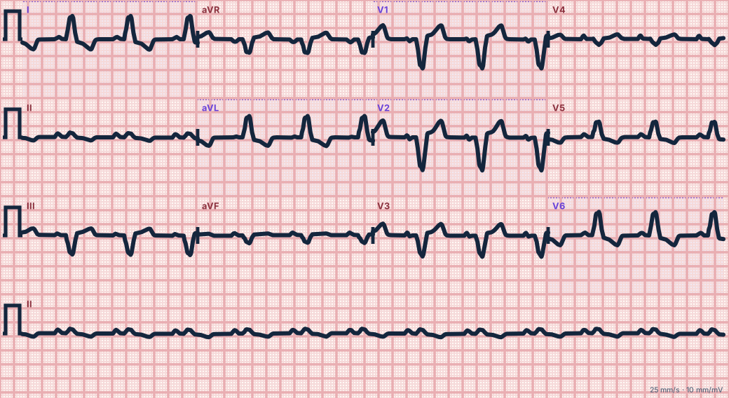
Scroll sideways to see the whole tracing, or select it to enlarge.
Expected discordance
In left bundle branch block the ST segment and T wave normally point opposite the main QRS deflection: elevation where the QRS is mostly negative (V1–V3) and depression where it is mostly positive (I, aVL, V5–V6).
The criteria
Concordant ST elevation of at least 1 mm in any lead with a positive QRS. Concordant ST depression of at least 1 mm in V1, V2 or V3. Excessively discordant ST elevation: in the modified criteria, ST elevation at least 25% of the depth of the preceding S wave (ST/S ratio ≤ −0.25).
Using them
Any one criterion makes acute occlusion likely in the right setting. The same approach applies to right ventricular paced rhythms. Absence of the criteria does not exclude occlusion.
How to read it
- Confirm left bundle branch block or ventricular pacing first.
- Check every lead for ST shifts in the same direction as the QRS.
- Measure ST elevation against the S wave depth in V1–V3.
Pitfalls
- A new left bundle branch block alone is no longer treated as a STEMI equivalent; use the criteria and the clinical picture.
Key numbers
- Concordant STE
- ≥ 1 mm, any lead
- Concordant STD
- ≥ 1 mm in V1–V3
- Excessive discordance
- ST/S ratio ≤ −0.25
Advanced Down to the ion channel
What the cells are doing
Pacemaker, atrial and ventricular action potentials, in step with the tracing above, with sodium, calcium and potassium moving through their channels.
Frequently asked questions
Occlusion in left bundle branch block: what is the key idea?
Left bundle branch block and ventricular pacing distort the ST segment by themselves, hiding acute occlusion. The Sgarbossa criteria, as modified by Smith, look for ST shifts that the block cannot explain.
Occlusion in left bundle branch block: what are the key numbers?
Concordant STE: ≥ 1 mm, any lead; Concordant STD: ≥ 1 mm in V1–V3; Excessive discordance: ST/S ratio ≤ −0.25.
Occlusion in left bundle branch block: how do you apply it to an ECG?
Confirm left bundle branch block or ventricular pacing first. Check every lead for ST shifts in the same direction as the QRS. Measure ST elevation against the S wave depth in V1–V3.
Occlusion in left bundle branch block: what are the common pitfalls?
A new left bundle branch block alone is no longer treated as a STEMI equivalent; use the criteria and the clinical picture.
Sources
This page does not list page-specific sources yet.
Every source the site cites, with links