
Electrophysiology
The cardiac vector
Why waves point up or down
At every instant the heart's electrical activity can be summed into one arrow, the cardiac vector. Each lead simply records how much of that arrow points toward it.
Last updated
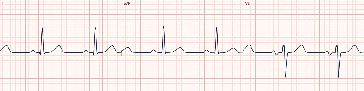
Example tracing, drawn by the lab's engine. Open the lab for a live, adjustable version.
At a glance
- Toward the lead
- Positive deflection
- Away from the lead
- Negative deflection
- Perpendicular
- Small or biphasic
Figures
Still images drawn by the lab's engine at 25 mm/s and 10 mm/mV, with the finding labeled. Select a figure to enlarge it.
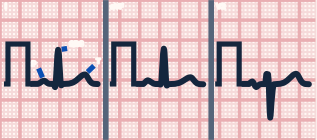
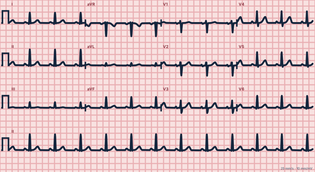
Scroll sideways to see the whole tracing, or select it to enlarge.
One arrow, many views
As the wavefront spreads, the vector changes direction and size. A lead records a positive deflection when the vector points toward its positive pole, negative when it points away and nothing when it points at right angles. That is why the same beat looks different in every lead.
The QRS loop
Tracing the tip of the vector through the QRS draws a loop: first the septum activates left to right, then the large left ventricular free wall pulls the vector down and to the left, and finally the base activates. Collapsing that loop onto each lead gives its QRS shape.
Mean vectors
Averaging the vector over the P wave, the QRS or the T wave gives the P, QRS and T axes. In the frontal plane they are read with the hexaxial system; in the horizontal plane with the chest leads.
How to read it
- Watch the vector arrow on the heart panel move through each beat.
- Switch to Chest leads 3D to see the same vector in three dimensions with its shadow on the chest plane.
Pitfalls
- The ECG records voltage at the skin, which is also shaped by distance, chest size and tissue between heart and electrode.
Key numbers
- Toward the lead
- Positive deflection
- Away from the lead
- Negative deflection
- Perpendicular
- Small or biphasic
Advanced Down to the ion channel
What the cells are doing
Pacemaker, atrial and ventricular action potentials, in step with the tracing above, with sodium, calcium and potassium moving through their channels.
Frequently asked questions
The cardiac vector: what is the key idea?
At every instant the heart's electrical activity can be summed into one arrow, the cardiac vector. Each lead simply records how much of that arrow points toward it.
The cardiac vector: what are the key numbers?
Toward the lead: Positive deflection; Away from the lead: Negative deflection; Perpendicular: Small or biphasic.
The cardiac vector: how do you apply it to an ECG?
Watch the vector arrow on the heart panel move through each beat. Switch to Chest leads 3D to see the same vector in three dimensions with its shadow on the chest plane.
The cardiac vector: what are the common pitfalls?
The ECG records voltage at the skin, which is also shaped by distance, chest size and tissue between heart and electrode.
Sources
This page does not list page-specific sources yet.
Every source the site cites, with links