
Electrophysiology
Cardiac action potentials
Ions, phases and the ECG
Every wave on the ECG is the sum of millions of single cells changing voltage. Pacemaker cells drift up to threshold on their own; working muscle cells fire fast and then hold a long plateau.
Last updated
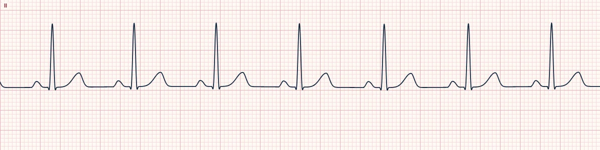
Example tracing, drawn by the lab's engine. Open the lab for a live, adjustable version.
At a glance
- Phase 0
- Sodium in: QRS
- Phase 2
- Calcium in: ST segment
- Phase 3
- Potassium out: T wave
- Pacemaker phase 4
- Funny current and calcium: slow rise to threshold
Figures
Still images drawn by the lab's engine at 25 mm/s and 10 mm/mV, with the finding labeled. Select a figure to enlarge it.
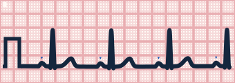
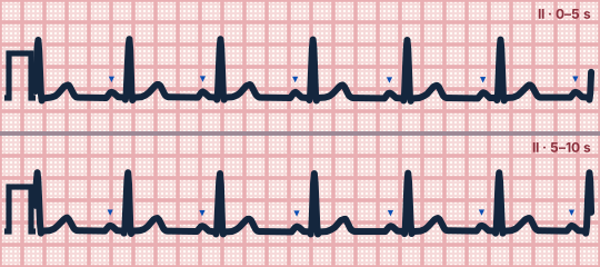
Scroll sideways to see the whole tracing, or select it to enlarge.
Ventricular muscle (phases 0–4)
Phase 0 is the rapid upstroke as sodium channels open, which on the ECG is the QRS. Phase 1 is a brief notch, phase 2 the plateau held by calcium entering (the ST segment), phase 3 repolarization as potassium leaves (the T wave) and phase 4 the stable resting potential.
Pacemaker cells
Sinus and AV node cells have no stable resting potential. In phase 4 the funny current (If) and calcium channels slowly depolarize them to threshold, and their upstroke is carried by calcium rather than sodium, which is why it is slower. Sympathetic stimulation steepens phase 4 and speeds the heart; vagal stimulation flattens it.
Refractory periods
During the plateau a cell cannot be re-excited. This protects the heart from tetany and is why a beat arriving very early is blocked or conducted with aberrancy.
How to read it
- Open the cells panel and watch the pacemaker, atrial and ventricular cells fire in step with the strip.
- Slow the speed to see sodium, calcium and potassium move through their channels.
Pitfalls
- Electrolyte changes act on these currents: high potassium flattens phase 0 and peaks the T wave; low calcium lengthens the plateau.
Key numbers
- Phase 0
- Sodium in: QRS
- Phase 2
- Calcium in: ST segment
- Phase 3
- Potassium out: T wave
- Pacemaker phase 4
- Funny current and calcium: slow rise to threshold
Advanced Down to the ion channel
What the cells are doing
Pacemaker, atrial and ventricular action potentials, in step with the tracing above, with sodium, calcium and potassium moving through their channels.
Frequently asked questions
Cardiac action potentials: what is the key idea?
Every wave on the ECG is the sum of millions of single cells changing voltage. Pacemaker cells drift up to threshold on their own; working muscle cells fire fast and then hold a long plateau.
Cardiac action potentials: what are the key numbers?
Phase 0: Sodium in: QRS; Phase 2: Calcium in: ST segment; Phase 3: Potassium out: T wave; Pacemaker phase 4: Funny current and calcium: slow rise to threshold.
Cardiac action potentials: how do you apply it to an ECG?
Open the cells panel and watch the pacemaker, atrial and ventricular cells fire in step with the strip. Slow the speed to see sodium, calcium and potassium move through their channels.
Cardiac action potentials: what are the common pitfalls?
Electrolyte changes act on these currents: high potassium flattens phase 0 and peaks the T wave; low calcium lengthens the plateau.
Sources
This page does not list page-specific sources yet.
Every source the site cites, with links