
Electrophysiology
The conduction system
From sinus node to Purkinje fibres
A chain of specialised tissue starts each beat, holds it at the AV node, then delivers it to both ventricles at almost the same moment. Each part has its own intrinsic rate and its own way of failing.
Last updated
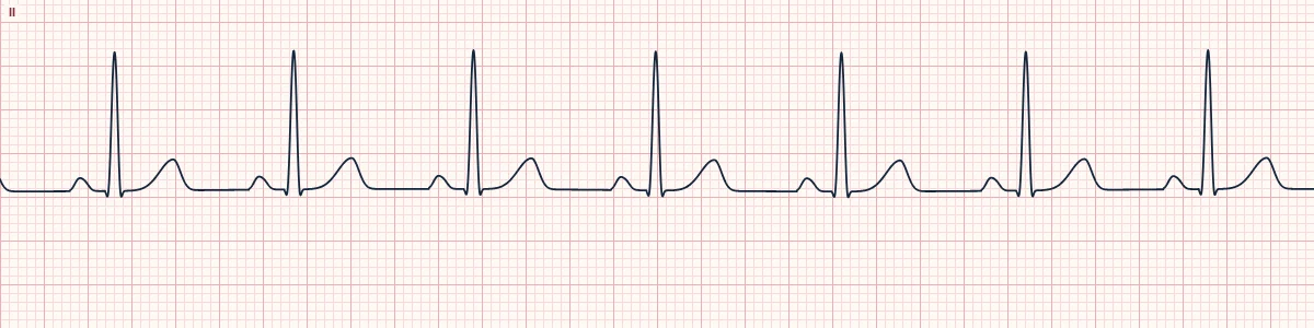
Example tracing, drawn by the lab's engine. Open the lab for a live, adjustable version.
At a glance
- Sinus node
- 60–100/min
- AV junction
- 40–60/min
- Ventricles
- 20–40/min
- AV node delay
- About 100 ms
Figures
Still images drawn by the lab's engine at 25 mm/s and 10 mm/mV, with the finding labeled. Select a figure to enlarge it.
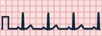
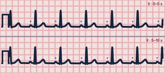
Scroll sideways to see the whole tracing, or select it to enlarge.
The pathway
The sinus node in the high right atrium fires; the impulse spreads through both atria and reaches the AV node, the only normal electrical connection to the ventricles. After a deliberate delay it passes through the bundle of His, divides into the right and left bundle branches (the left splitting into anterior and posterior fascicles) and reaches the Purkinje network lining the ventricles.
Backup pacemakers
Every part of the system can generate its own impulses, but more slowly the further down it sits. Normally the sinus node is fastest and resets the others; if it fails, the next one down takes over as an escape rhythm.
Where blocks happen
Sinus node disease gives pauses and bradycardia. AV node block gives a long PR and Wenckebach. Block below the AV node (in the His bundle or both bundle branches) gives Mobitz II and complete heart block with slow, wide escape rhythms.
How to read it
- Watch the impulse travel the pathway on the conduction heart in step with each wave on the strip.
- Match each part of the PQRST to the part of the pathway that is active.
Pitfalls
- The AV node is not a pacemaker in health; the junction only paces when the sinus node fails or slows.
Key numbers
- Sinus node
- 60–100/min
- AV junction
- 40–60/min
- Ventricles
- 20–40/min
- AV node delay
- About 100 ms
Advanced Down to the ion channel
What the cells are doing
Pacemaker, atrial and ventricular action potentials, in step with the tracing above, with sodium, calcium and potassium moving through their channels.
Frequently asked questions
The conduction system: what is the key idea?
A chain of specialised tissue starts each beat, holds it at the AV node, then delivers it to both ventricles at almost the same moment. Each part has its own intrinsic rate and its own way of failing.
The conduction system: what are the key numbers?
Sinus node: 60–100/min; AV junction: 40–60/min; Ventricles: 20–40/min; AV node delay: About 100 ms.
The conduction system: how do you apply it to an ECG?
Watch the impulse travel the pathway on the conduction heart in step with each wave on the strip. Match each part of the PQRST to the part of the pathway that is active.
The conduction system: what are the common pitfalls?
The AV node is not a pacemaker in health; the junction only paces when the sinus node fails or slows.
Sources
This page does not list page-specific sources yet.
Every source the site cites, with links