
Position & lead placement
Dextrocardia
The heart lies on the right side of the chest as a mirror image. Standard lead placement then records the frontal plane reversed, with shrinking chest-lead complexes.
Last updated
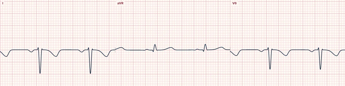
Example tracing, drawn by the lab's engine. Open the lab for a live, adjustable version.
At a glance
- Lead I
- P wave, QRS and T wave all inverted
- aVR
- Upright complexes (aVR and aVL appear swapped)
- Axis
- Right axis deviation
- V1–V6
- R wave shrinks from V1 to V6 (reversed progression)
Figures
Still images drawn by the lab's engine at 25 mm/s and 10 mm/mV, with the finding labeled. Select a figure to enlarge it.
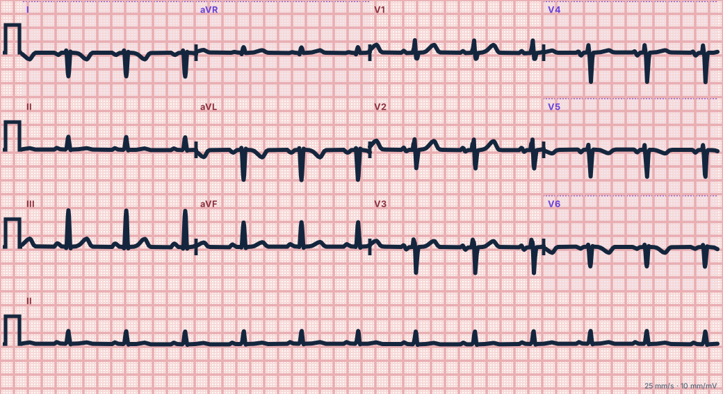
Scroll sideways to see the whole tracing, or select it to enlarge.
What the heart is doing
Activation spreads through a normal conduction system but from left to right across the chest. Lead I sees it moving away, and the left-sided chest electrodes sit far from the heart.
Why it looks this way
This is dextrocardia because lead I is globally inverted with an upright aVR, and the R wave shrinks progressively from V1 to V6.
Why it matters
Usually harmless when all organs are reversed. The practical point is to record a right-sided ECG for interpretation, and to place defibrillation pads to match the heart's position.
What to look for
- Everything in lead I is upside down, including the P wave.
- The chest leads get smaller toward V6 because they move away from the heart.
- Repeat with the chest leads on the right side and arm leads swapped: the ECG normalizes.
Criteria
- Lead I
- P wave, QRS and T wave all inverted
- aVR
- Upright complexes (aVR and aVL appear swapped)
- Axis
- Right axis deviation
- V1–V6
- R wave shrinks from V1 to V6 (reversed progression)
Causes
- Congenital mirror-image position of the heart
- Complete reversal of the organs (situs inversus)
- Primary ciliary dyskinesia
- Associated congenital heart defects
Can be mistaken for
- Arm lead reversal — the same limb-lead picture but normal chest leads Compare
- Lateral infarction — Q waves in I and aVL with an upright P wave
- Right ventricular hypertrophy — right axis with a tall R in V1 Compare
Advanced Down to the ion channel
What the cells are doing
Pacemaker, atrial and ventricular action potentials, in step with the tracing above, with sodium, calcium and potassium moving through their channels.
Frequently asked questions
What are the ECG criteria for dextrocardia?
Lead I: P wave, QRS and T wave all inverted. aVR: upright complexes (aVR and aVL appear swapped). Axis: right axis deviation. V1–V6: R wave shrinks from V1 to V6 (reversed progression).
How is dextrocardia identified on an ECG?
This is dextrocardia because lead I is globally inverted with an upright aVR, and the R wave shrinks progressively from V1 to V6.
What can dextrocardia be mistaken for?
Arm lead reversal: the same limb-lead picture but normal chest leads. Lateral infarction: Q waves in I and aVL with an upright P wave. Right ventricular hypertrophy: right axis with a tall R in V1.
What causes dextrocardia?
Common causes: congenital mirror-image position of the heart; complete reversal of the organs (situs inversus); primary ciliary dyskinesia; and associated congenital heart defects.
Sources
This page does not list page-specific sources yet.
Every source the site cites, with links