
Other conditions
Draft — pending owner review
Atrial septal defect (crochetage sign)
A notch near the peak of the R wave in the inferior leads, linked to secundum atrial septal defect. With an incomplete RBBB pattern and a rightward axis it strongly suggests the defect.
Last updated
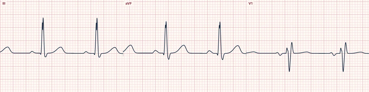
Example tracing, drawn by the lab's engine. Open the lab for a live, adjustable version.
At a glance
- Notch
- Near the apex of the R wave in II, III or aVF
- Specificity
- Highest when present in all three inferior leads or with incomplete RBBB
- V1
- rSr′ from right ventricular volume load
- Axis
- Rightward
Figures
Still images drawn by the lab's engine at 25 mm/s and 10 mm/mV, with the finding labeled. Select a figure to enlarge it.
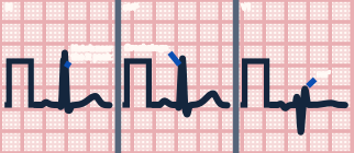
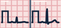
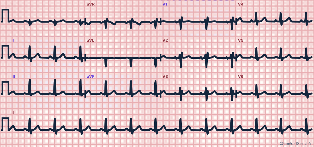
Scroll sideways to see the whole tracing, or select it to enlarge.
What the heart is doing
The notch is not fully explained; it is thought to reflect altered activation around the enlarged right heart and septum. Right ventricular volume overload delays right-sided activation, giving the r′ in V1.
Why it looks this way
This is the crochetage sign because the R waves in the inferior leads carry a notch near their peak, with an rSr′ in V1 and a rightward axis.
Why it matters
Atrial septal defects often go unnoticed into adult life and present with breathlessness, atrial arrhythmias or paradoxical embolism. A crochetage notch is a cheap clue that should prompt an echocardiogram.
What to look for
- Zoom in on the tip of the R wave in II, III and aVF: a small notch, like a crochet hook.
- Check V1 for the rSr′ of right ventricular volume load.
- Listen for a fixed split second heart sound and arrange an echocardiogram.
Criteria
- Notch
- Near the apex of the R wave in II, III or aVF
- Specificity
- Highest when present in all three inferior leads or with incomplete RBBB
- V1
- rSr′ from right ventricular volume load
- Axis
- Rightward
- Rhythm
- Atrial fibrillation or flutter in older adults
Causes
- Ostium secundum atrial septal defect
- Patent foramen ovale (less often)
- Ventricular septal defect (less often)
Can be mistaken for
- Incomplete right bundle branch block — rSr′ without inferior R-wave notching Compare
- Right ventricular hypertrophy — dominant R wave in V1 and right axis deviation Compare
- Fragmented QRS from scar — notching in a territory with Q waves
Advanced Down to the ion channel
What the cells are doing
Pacemaker, atrial and ventricular action potentials, in step with the tracing above, with sodium, calcium and potassium moving through their channels.
Frequently asked questions
What are the ECG criteria for atrial septal defect (crochetage sign)?
Notch: near the apex of the R wave in II, III or aVF. Specificity: highest when present in all three inferior leads or with incomplete RBBB. V1: rSr′ from right ventricular volume load. Axis: rightward. Rhythm: atrial fibrillation or flutter in older adults.
How is atrial septal defect (crochetage sign) identified on an ECG?
This is the crochetage sign because the R waves in the inferior leads carry a notch near their peak, with an rSr′ in V1 and a rightward axis.
What can atrial septal defect (crochetage sign) be mistaken for?
Incomplete right bundle branch block: rSr′ without inferior R-wave notching. Right ventricular hypertrophy: dominant R wave in V1 and right axis deviation. Fragmented QRS from scar: notching in a territory with Q waves.
What causes atrial septal defect (crochetage sign)?
Common causes: ostium secundum atrial septal defect; patent foramen ovale (less often); and ventricular septal defect (less often).
Sources
This page does not list page-specific sources yet.
Every source the site cites, with links