
Repolarization & channelopathies
Draft — pending owner review
Brugada pattern, type 2 (saddleback)
The saddleback Brugada pattern: a high J point in V1–V2, an ST segment that dips but stays raised, then an upright T wave. It is suggestive only; Brugada syndrome needs the type 1 pattern.
Last updated
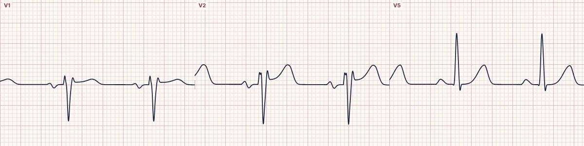
Example tracing, drawn by the lab's engine. Open the lab for a live, adjustable version.
At a glance
- V1–V2
- Saddleback ST elevation
- J point
- High take-off, usually ≥ 2 mm
- ST trough
- Stays ≥ 0.5 mm above the baseline
- T wave
- Upright in V2, variable in V1
Figures
Still images drawn by the lab's engine at 25 mm/s and 10 mm/mV, with the finding labeled. Select a figure to enlarge it.
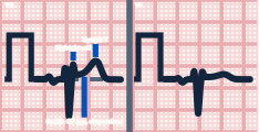
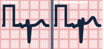
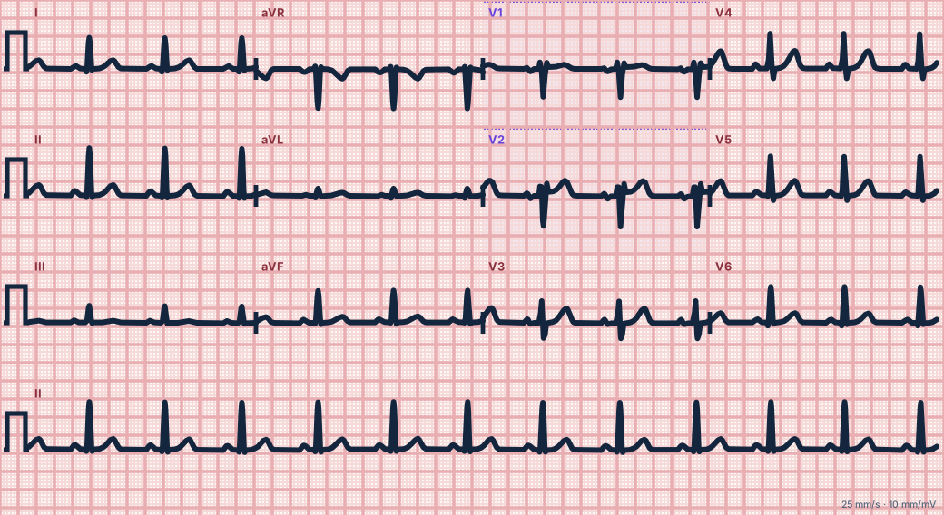
Scroll sideways to see the whole tracing, or select it to enlarge.
What the heart is doing
A milder imbalance between inward sodium and outward potassium currents in the right ventricular outflow epicardium raises the J point without reversing repolarization, so the T wave stays upright.
Why it looks this way
This is a type 2 Brugada pattern because V2 shows a high J-point take-off, a raised ST trough and an upright T wave in a saddleback shape.
Why it matters
A type 2 pattern alone does not establish Brugada syndrome. It calls for a careful history (syncope, nocturnal agonal breathing, family sudden death), repeat ECGs with high leads and expert referral. Treat fever promptly.
What to look for
- Trace V2: a high take-off, a dip, then an upright T wave, like a saddle.
- Look for an r′ in V1–V2 without a wide S wave in I and V6.
- Record V1–V2 one and two rib spaces higher: a type 1 pattern may appear.
Criteria
- V1–V2
- Saddleback ST elevation
- J point
- High take-off, usually ≥ 2 mm
- ST trough
- Stays ≥ 0.5 mm above the baseline
- T wave
- Upright in V2, variable in V1
- Diagnosis
- Needs conversion to type 1, spontaneously, with fever or on drug challenge
Causes
- Inherited sodium-channel variants
- Fever
- Sodium-channel blocking drugs
- Athletic or normal variant
- V1–V2 placed too high
Can be mistaken for
- Brugada pattern — type 1 is coved, sloping into an inverted T wave Compare
- Incomplete right bundle branch block — a narrow r′ with no ST elevation Compare
- Athlete's heart — benign J-point elevation, usually most marked in V2–V4
Advanced Down to the ion channel
What the cells are doing
Pacemaker, atrial and ventricular action potentials, in step with the tracing above, with sodium, calcium and potassium moving through their channels.
Frequently asked questions
What are the ECG criteria for brugada pattern, type 2 (saddleback)?
V1–V2: saddleback ST elevation. J point: high take-off, usually ≥ 2 mm. ST trough: stays ≥ 0.5 mm above the baseline. T wave: upright in V2, variable in V1. Diagnosis: needs conversion to type 1, spontaneously, with fever or on drug challenge.
How is brugada pattern, type 2 (saddleback) identified on an ECG?
This is a type 2 Brugada pattern because V2 shows a high J-point take-off, a raised ST trough and an upright T wave in a saddleback shape.
What can brugada pattern, type 2 (saddleback) be mistaken for?
Brugada pattern: type 1 is coved, sloping into an inverted T wave. Incomplete right bundle branch block: a narrow r′ with no ST elevation. Athlete's heart: benign J-point elevation, usually most marked in V2–V4.
What causes brugada pattern, type 2 (saddleback)?
Common causes: inherited sodium-channel variants; fever; sodium-channel blocking drugs; athletic or normal variant; and V1–V2 placed too high.
Sources
This page does not list page-specific sources yet.
Every source the site cites, with links