
Chamber enlargement
Left ventricular hypertrophy with strain
Thickened left ventricular muscle produces abnormally large QRS voltages. The strain pattern adds ST depression and T inversion in the lateral leads.
Last updated
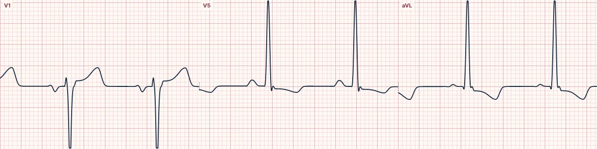
Example tracing, drawn by the lab's engine. Open the lab for a live, adjustable version.
At a glance
- Voltage
- S in V1 + R in V5 or V6 ≥ 35 mm, or R in aVL ≥ 11 mm
- ST–T (strain)
- Downsloping ST depression and asymmetric T inversion in I, aVL, V5–V6
- V1–V3
- ST elevation proportional to the deep S wave
- Axis
- Normal or leftward
Figures
Still images drawn by the lab's engine at 25 mm/s and 10 mm/mV, with the finding labeled. Select a figure to enlarge it.
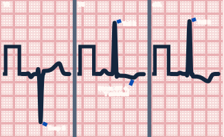
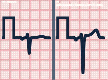
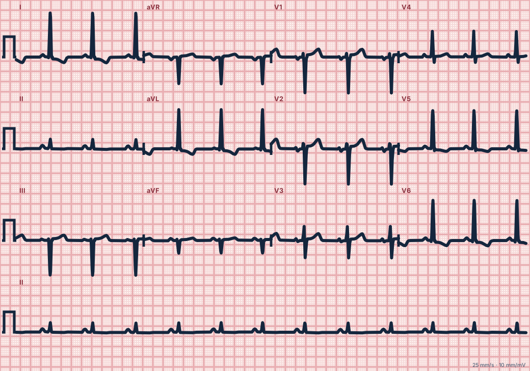
Scroll sideways to see the whole tracing, or select it to enlarge.
What the heart is doing
More left ventricular muscle generates larger leftward, posterior forces and takes slightly longer to activate. Repolarization of the thick wall is reversed, turning the ST–T away from the QRS.
Why it looks this way
This is LVH with strain because voltage criteria are met and the lateral leads show downsloping ST depression with asymmetric T inversion.
Why it matters
A marker of chronic pressure or volume load and a higher risk of heart failure, arrhythmia and ischemia. Strain changes can imitate or hide ischemia, so compare with previous ECGs.
What to look for
- Add the deepest S in V1 to the tallest R in V5 or V6: 35 mm or more suggests LVH.
- Strain T waves slope down slowly and return quickly; ischemic T waves are symmetric.
- Expect matching left atrial enlargement.
Criteria
- Voltage
- S in V1 + R in V5 or V6 ≥ 35 mm, or R in aVL ≥ 11 mm
- ST–T (strain)
- Downsloping ST depression and asymmetric T inversion in I, aVL, V5–V6
- V1–V3
- ST elevation proportional to the deep S wave
- Axis
- Normal or leftward
- QRS
- Slightly prolonged (< 120 ms)
Causes
- Long-standing hypertension
- Aortic stenosis
- Aortic or mitral regurgitation
- Hypertrophic cardiomyopathy
- Athletic training (voltage without strain)
Can be mistaken for
Advanced Down to the ion channel
What the cells are doing
Pacemaker, atrial and ventricular action potentials, in step with the tracing above, with sodium, calcium and potassium moving through their channels.
Frequently asked questions
What are the ECG criteria for left ventricular hypertrophy with strain?
Voltage: S in V1 + R in V5 or V6 ≥ 35 mm, or R in aVL ≥ 11 mm. ST–T (strain): downsloping ST depression and asymmetric T inversion in I, aVL, V5–V6. V1–V3: ST elevation proportional to the deep S wave. Axis: normal or leftward. QRS: slightly prolonged (< 120 ms).
How is left ventricular hypertrophy with strain identified on an ECG?
This is LVH with strain because voltage criteria are met and the lateral leads show downsloping ST depression with asymmetric T inversion.
What can left ventricular hypertrophy with strain be mistaken for?
LBBB: QRS 120 ms or more with no septal q waves. Lateral ischemia: horizontal ST depression or symmetric T inversion, changing over time. Anterior STEMI: V1–V3 elevation out of proportion to the S wave.
What causes left ventricular hypertrophy with strain?
Common causes: long-standing hypertension; aortic stenosis; aortic or mitral regurgitation; hypertrophic cardiomyopathy; and athletic training (voltage without strain).
Sources
This page does not list page-specific sources yet.
Every source the site cites, with links