
Conduction system disease
Draft — pending owner review
Lenègre–Lev disease
Progressive fibrosis of the conduction system
Degenerative fibrosis and calcification of both bundle branches, progressing over years from bundle branch block to bifascicular block and finally complete heart block. It is a leading cause of acquired heart block in older adults.
Two descriptions, one process
Lenègre described slow fibrosis of the bundle branches in middle-aged people; Lev described sclerosis and calcification of the fibrous skeleton around the conduction system in older people. Both destroy the bundle branches rather than the AV node.
What the ECG shows over time
RBBB or LBBB, then an added fascicular block, sometimes a long PR, then intermittent Mobitz II or complete heart block with a slow, wide escape rhythm. Fainting from sudden complete block (Stokes–Adams attacks) may be the first symptom.
Why it matters
Because the block is below the AV node, escape rhythms are slow and unreliable and atropine is unlikely to help. Pacing is the definitive treatment.
About the demonstration
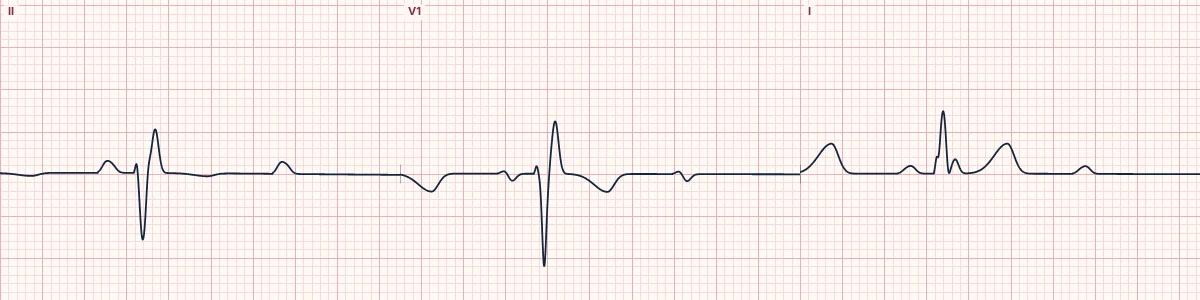
The tracing shows one stage of the disease: Mobitz II block in a patient with RBBB and left anterior fascicular block.
How to read it
- Compare serial ECGs for accumulating conduction defects.
- In any bundle branch block with syncope, look for dropped beats on a long strip.
- Note the escape QRS width in complete block: wide means a low, unreliable escape.
Pitfalls
- A bundle branch block stable for years can still progress.
- Atropine speeds the sinus node but does not help block below the AV node and can make the block ratio worse.
Frequently asked questions
Lenègre–Lev disease: what is the key idea?
Degenerative fibrosis and calcification of both bundle branches, progressing over years from bundle branch block to bifascicular block and finally complete heart block. It is a leading cause of acquired heart block in older adults.
Lenègre–Lev disease: what are the key numbers?
Level of block: Below the AV node (bundle branches); Typical patient: Older adult, often with hypertension or coronary disease; Presentation: Syncope, fatigue, slow wide escape rhythm; Treatment: Permanent pacing.
Lenègre–Lev disease: how do you apply it to an ECG?
Compare serial ECGs for accumulating conduction defects. In any bundle branch block with syncope, look for dropped beats on a long strip. Note the escape QRS width in complete block: wide means a low, unreliable escape.
Lenègre–Lev disease: what are the common pitfalls?
A bundle branch block stable for years can still progress. Atropine speeds the sinus node but does not help block below the AV node and can make the block ratio worse.