
AV blocks
Second-degree AV block, Mobitz II
Some P waves suddenly fail to conduct with no warning lengthening of the PR. The block lies below the AV node and can progress abruptly to complete block.
Last updated
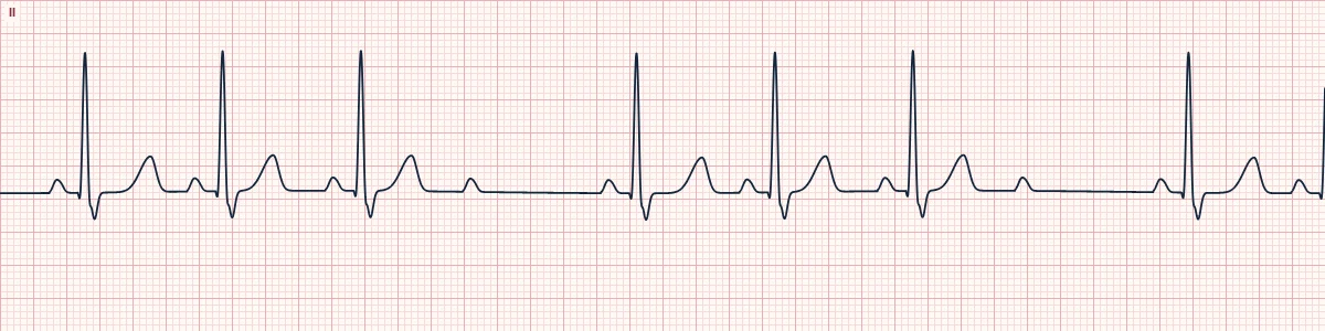
Example tracing, drawn by the lab's engine. Open the lab for a live, adjustable version.
At a glance
- Rate
- Atrial regular; ventricular slower
- Rhythm
- Irregular because of dropped beats
- P waves
- Regular; some not followed by a QRS
- PR interval
- Constant on all conducted beats
Figures
Still images drawn by the lab's engine at 25 mm/s and 10 mm/mV, with the finding labeled. Select a figure to enlarge it.
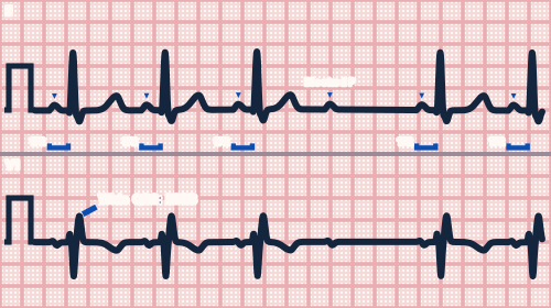
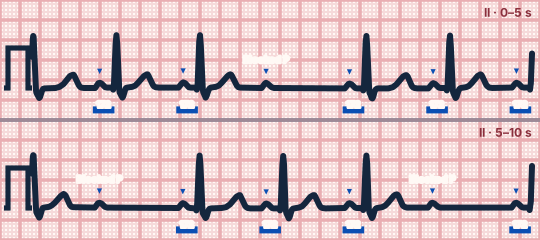
Scroll sideways to see the whole tracing, or select it to enlarge.
What the heart is doing
The block is in the His bundle or bundle branches, which conduct all-or-nothing. Impulses pass the AV node normally, so the PR is constant, but some are stopped completely below it.
Why it looks this way
This is Mobitz II because P waves are intermittently not conducted while the PR of the conducted beats stays constant.
Why it matters
Unstable and unpredictable: it can jump to complete block or ventricular standstill. Atropine is often ineffective; prepare for pacing. A permanent pacemaker is usually needed.
What to look for
- Compare the PR before and after the dropped beat: they are identical.
- March the P waves through the pause; the non-conducted P is exactly on time.
- The pause equals two normal P–P intervals.
Criteria
- Rate
- Atrial regular; ventricular slower
- Rhythm
- Irregular because of dropped beats
- P waves
- Regular; some not followed by a QRS
- PR interval
- Constant on all conducted beats
- QRS
- Often wide (bundle branch block); may be narrow
Causes
- Anterior myocardial infarction
- Degenerative fibrosis of the conduction system
- Cardiac surgery or valve procedures
- Myocarditis
- Infiltrative disease (sarcoidosis, amyloid)
Can be mistaken for
- Mobitz I — the PR is longer before the dropped beat than after it Compare
- Non-conducted PACs — the blocked P wave arrives early
- 2:1 AV block — every other P wave is blocked, so the type cannot be judged Compare
Advanced Down to the ion channel
What the cells are doing
Pacemaker, atrial and ventricular action potentials, in step with the tracing above, with sodium, calcium and potassium moving through their channels.
Frequently asked questions
What are the ECG criteria for second-degree AV block, Mobitz II?
Rate: atrial regular; ventricular slower. Rhythm: irregular because of dropped beats. P waves: regular; some not followed by a QRS. PR interval: constant on all conducted beats. QRS: often wide (bundle branch block); may be narrow.
How is second-degree AV block, Mobitz II identified on an ECG?
This is Mobitz II because P waves are intermittently not conducted while the PR of the conducted beats stays constant.
What can second-degree AV block, Mobitz II be mistaken for?
Mobitz I: the PR is longer before the dropped beat than after it. Non-conducted PACs: the blocked P wave arrives early. 2:1 AV block: every other P wave is blocked, so the type cannot be judged.
What causes second-degree AV block, Mobitz II?
Common causes: anterior myocardial infarction; degenerative fibrosis of the conduction system; cardiac surgery or valve procedures; myocarditis; and infiltrative disease (sarcoidosis, amyloid).
Practise it in the ACLS Lab
Sources
This page does not list page-specific sources yet.
Every source the site cites, with links