
Ventricular
Non-sustained ventricular tachycardia
A short burst of ventricular tachycardia: three or more ventricular beats in a row faster than 100/min that stops by itself within 30 seconds.
Last updated
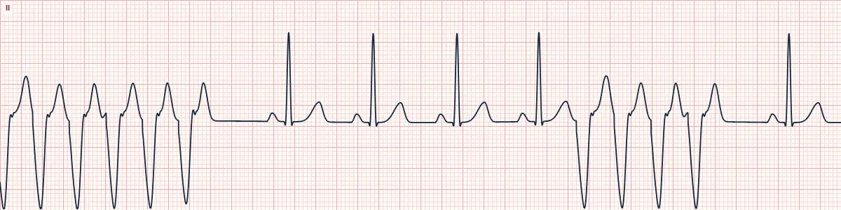
Example tracing, drawn by the lab's engine. Open the lab for a live, adjustable version.
At a glance
- Rate
- > 100/min during the run (here about 170)
- Duration
- ≥ 3 beats, ending within 30 seconds
- Rhythm
- Regular during the run
- P waves
- None related to the wide beats
Figures
Still images drawn by the lab's engine at 25 mm/s and 10 mm/mV, with the finding labeled. Select a figure to enlarge it.
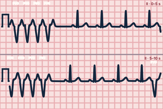
Scroll sideways to see the whole tracing, or select it to enlarge.
What the heart is doing
A re-entry circuit around scar, or a rapidly firing ventricular focus, takes over for a few beats. It then extinguishes spontaneously and the sinus node resumes control.
Why it looks this way
This is non-sustained VT because three or more consecutive wide ventricular beats run faster than 100/min and stop spontaneously in under 30 seconds.
Why it matters
A warning sign, especially with structural heart disease, where it predicts sustained VT and sudden death. Correct electrolytes and ischemia, monitor continuously and arrange cardiac evaluation.
What to look for
- A sudden salvo of wide beats that interrupts sinus rhythm and then stops spontaneously.
- Count the beats: three or more consecutive ventricular beats.
- Note the patient's symptoms and pulse during the run.
Criteria
- Rate
- > 100/min during the run (here about 170)
- Duration
- ≥ 3 beats, ending within 30 seconds
- Rhythm
- Regular during the run
- P waves
- None related to the wide beats
- QRS
- Wide (≥ 120 ms), uniform during the run
Causes
- Myocardial ischemia or prior infarction
- Cardiomyopathy and heart failure
- Hypokalemia, hypomagnesemia
- QT-prolonging drugs
- Stimulants
- Inherited arrhythmia syndromes
Can be mistaken for
- SVT with aberrancy — starts with an early P wave and has a typical bundle branch shape
- Artifact — sinus QRS complexes can be marched through the 'run'
- Accelerated idioventricular rhythm — wide run at under about 110/min Compare
Advanced Down to the ion channel
What the cells are doing
Pacemaker, atrial and ventricular action potentials, in step with the tracing above, with sodium, calcium and potassium moving through their channels.
Frequently asked questions
What are the ECG criteria for non-sustained ventricular tachycardia?
Rate: > 100/min during the run (here about 170). Duration: ≥ 3 beats, ending within 30 seconds. Rhythm: regular during the run. P waves: none related to the wide beats. QRS: wide (≥ 120 ms), uniform during the run.
How is non-sustained ventricular tachycardia identified on an ECG?
This is non-sustained VT because three or more consecutive wide ventricular beats run faster than 100/min and stop spontaneously in under 30 seconds.
What can non-sustained ventricular tachycardia be mistaken for?
SVT with aberrancy: starts with an early P wave and has a typical bundle branch shape. Artifact: sinus QRS complexes can be marched through the 'run'. Accelerated idioventricular rhythm: wide run at under about 110/min.
What causes non-sustained ventricular tachycardia?
Common causes: myocardial ischemia or prior infarction; cardiomyopathy and heart failure; hypokalemia, hypomagnesemia; QT-prolonging drugs; stimulants; and inherited arrhythmia syndromes.
Sources
This page does not list page-specific sources yet.
Every source the site cites, with links