
Ventricular
Ventricular couplets
Two PVCs back to back. They show a more irritable ventricle than single PVCs and sit one beat short of ventricular tachycardia.
Last updated
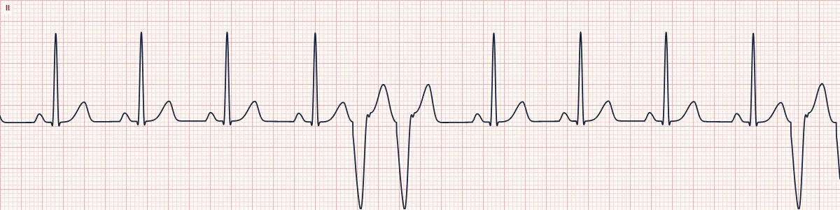
Example tracing, drawn by the lab's engine. Open the lab for a live, adjustable version.
At a glance
- Rate
- That of the underlying rhythm
- Rhythm
- Irregular because of paired early beats
- P waves
- None before the ventricular beats
- QRS
- Two consecutive wide (≥ 120 ms) beats
Figures
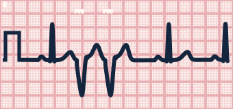
Still images drawn by the lab's engine at 25 mm/s and 10 mm/mV, with the finding labeled. Select a figure to enlarge it.
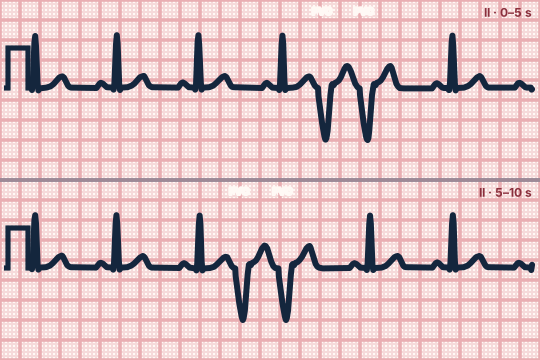
Scroll sideways to see the whole tracing, or select it to enlarge.
What the heart is doing
A ventricular focus or small re-entry circuit fires twice in succession before the sinus node regains control. Both beats spread slowly through muscle, giving wide complexes.
Why it looks this way
These are ventricular couplets because two wide premature ventricular beats occur consecutively, followed by a pause and a return to sinus rhythm.
Why it matters
Higher-grade ectopy than isolated PVCs. With ischemia, heart failure or electrolyte loss it predicts runs of VT; monitor closely and correct the cause.
What to look for
- Two wide, bizarre beats in a row, then a pause before sinus rhythm resumes.
- Count carefully: two is a couplet, three or more is ventricular tachycardia.
- Here the PVCs are upright in V1 and negative in the inferior leads, pointing to a left ventricular focus.
Criteria
- Rate
- That of the underlying rhythm
- Rhythm
- Irregular because of paired early beats
- P waves
- None before the ventricular beats
- QRS
- Two consecutive wide (≥ 120 ms) beats
- Pause
- Follows the second PVC
Causes
- Myocardial ischemia or infarction
- Cardiomyopathy and heart failure
- Hypokalemia, hypomagnesemia
- Hypoxia
- Stimulants
- QT-prolonging drugs
Can be mistaken for
- Non-sustained VT — three or more ventricular beats in a row Compare
- Two aberrantly conducted PACs — each wide beat is preceded by a P wave
- Artifact — the underlying QRS complexes march through undisturbed
Advanced Down to the ion channel
What the cells are doing
Pacemaker, atrial and ventricular action potentials, in step with the tracing above, with sodium, calcium and potassium moving through their channels.
Frequently asked questions
What are the ECG criteria for ventricular couplets?
Rate: that of the underlying rhythm. Rhythm: irregular because of paired early beats. P waves: none before the ventricular beats. QRS: two consecutive wide (≥ 120 ms) beats. Pause: follows the second PVC.
How is ventricular couplets identified on an ECG?
These are ventricular couplets because two wide premature ventricular beats occur consecutively, followed by a pause and a return to sinus rhythm.
What can ventricular couplets be mistaken for?
Non-sustained VT: three or more ventricular beats in a row. Two aberrantly conducted PACs: each wide beat is preceded by a P wave. Artifact: the underlying QRS complexes march through undisturbed.
What causes ventricular couplets?
Common causes: myocardial ischemia or infarction; cardiomyopathy and heart failure; hypokalemia, hypomagnesemia; hypoxia; stimulants; and QT-prolonging drugs.
Sources
This page does not list page-specific sources yet.
Every source the site cites, with links