
Ventricular
Idioventricular rhythm
A slow escape rhythm from the ventricles at 20–40/min, appearing when all higher pacemakers have failed. Wide QRS, no P waves.
Last updated
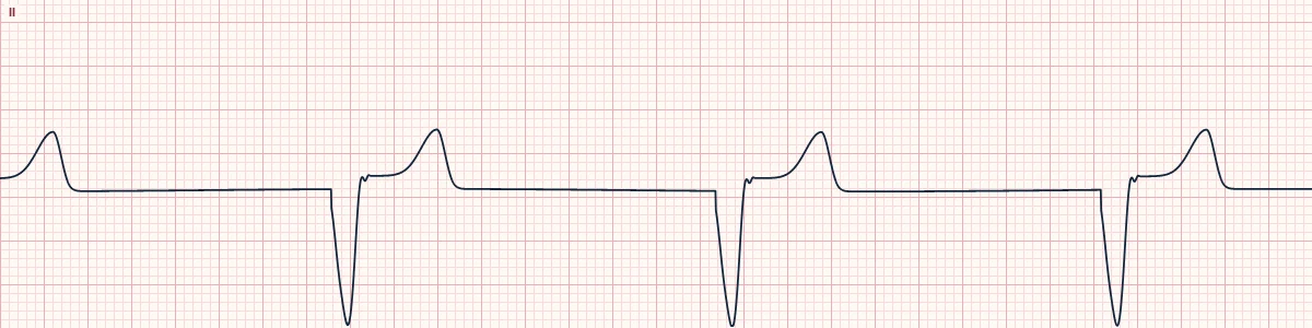
Example tracing, drawn by the lab's engine. Open the lab for a live, adjustable version.
At a glance
- Rate
- 20–40/min
- Rhythm
- Regular
- P waves
- Absent
- PR interval
- None
Figures
Still images drawn by the lab's engine at 25 mm/s and 10 mm/mV, with the finding labeled. Select a figure to enlarge it.
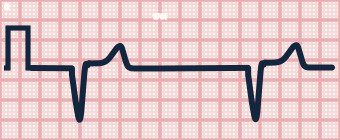
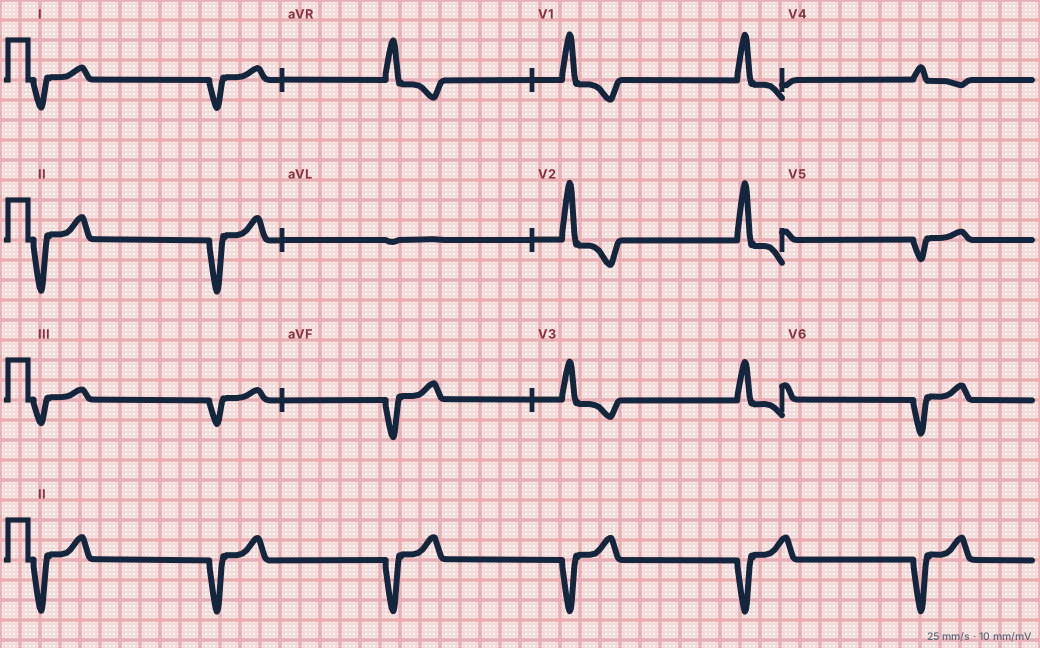
Scroll sideways to see the whole tracing, or select it to enlarge.
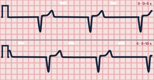
Scroll sideways to see the whole tracing, or select it to enlarge.
What the heart is doing
With no impulse from the sinus node or AV junction, a ventricular focus fires at its slow intrinsic rate. Activation spreads muscle to muscle, producing a wide QRS.
Why it looks this way
This is an idioventricular rhythm because it is regular at 20–40/min with wide QRS complexes and no P waves.
Why it matters
The heart's last backup pacemaker. Never suppress it with antiarrhythmics, which could cause asystole. If there is a pulse, support the rate and prepare for pacing; if not, treat as cardiac arrest.
What to look for
- Very slow, wide, regular complexes with large T waves pointing the opposite way.
- No P waves anywhere on the strip.
- Check for a pulse immediately; this rhythm is often pulseless.
Criteria
- Rate
- 20–40/min
- Rhythm
- Regular
- P waves
- Absent
- PR interval
- None
- QRS
- Wide (≥ 120 ms), bizarre
Causes
- Failure of the sinus node and junction
- Complete AV block
- Post-arrest and dying heart
- Severe hypoxia or acidosis
- Hyperkalemia
- Drug toxicity
Can be mistaken for
- Third-degree AV block with ventricular escape — P waves are present but unrelated Compare
- Junctional escape with bundle branch block — rate 40–60/min
- Agonal rhythm — slower, irregular, with deteriorating complexes Compare
Advanced Down to the ion channel
What the cells are doing
Pacemaker, atrial and ventricular action potentials, in step with the tracing above, with sodium, calcium and potassium moving through their channels.
Frequently asked questions
What are the ECG criteria for idioventricular rhythm?
Rate: 20–40/min. Rhythm: regular. P waves: absent. PR interval: none. QRS: wide (≥ 120 ms), bizarre.
How is idioventricular rhythm identified on an ECG?
This is an idioventricular rhythm because it is regular at 20–40/min with wide QRS complexes and no P waves.
What can idioventricular rhythm be mistaken for?
Third-degree AV block with ventricular escape: P waves are present but unrelated. Junctional escape with bundle branch block: rate 40–60/min. Agonal rhythm: slower, irregular, with deteriorating complexes.
What causes idioventricular rhythm?
Common causes: failure of the sinus node and junction; complete AV block; post-arrest and dying heart; severe hypoxia or acidosis; hyperkalemia; and drug toxicity.
Practise it in the ACLS Lab
Sources
This page does not list page-specific sources yet.
Every source the site cites, with links