
Ventricular
Premature ventricular complexes
Early beats that start in the ventricular muscle. They spread slowly from cell to cell, so the QRS is wide and oddly shaped with no P wave in front.
Last updated
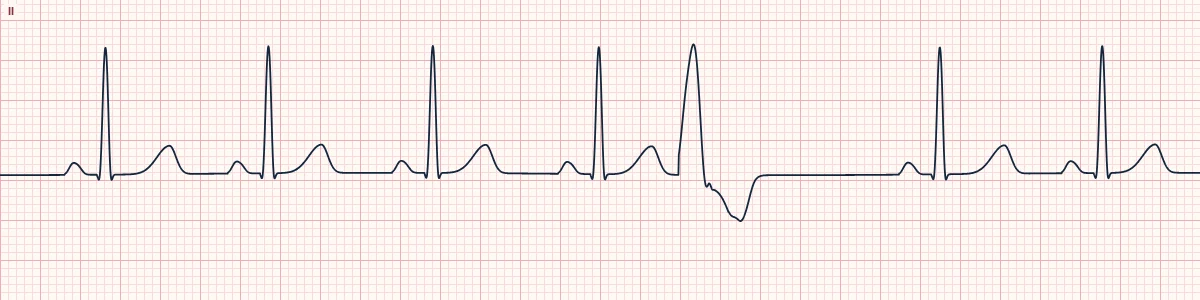
Example tracing, drawn by the lab's engine. Open the lab for a live, adjustable version.
At a glance
- Rate
- That of the underlying rhythm
- Rhythm
- Irregular because of early beats
- P waves
- None before the early beat
- QRS
- Wide (≥ 120 ms), bizarre, premature
Figures
Still images drawn by the lab's engine at 25 mm/s and 10 mm/mV, with the finding labeled. Select a figure to enlarge it.
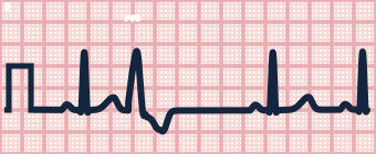
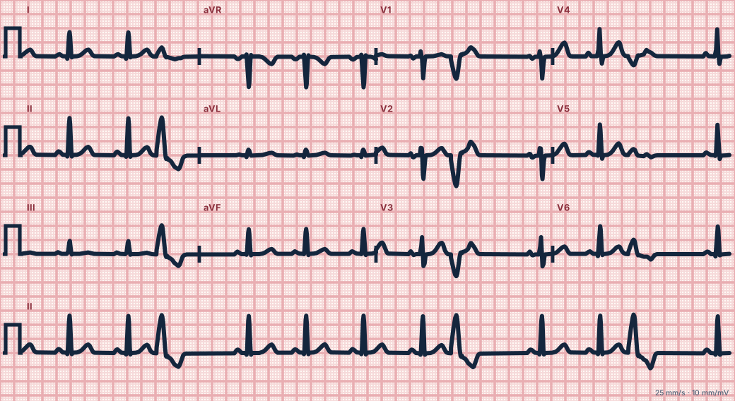
Scroll sideways to see the whole tracing, or select it to enlarge.
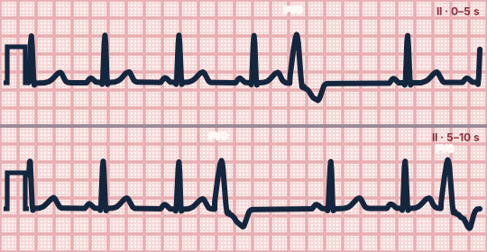
Scroll sideways to see the whole tracing, or select it to enlarge.
What the heart is doing
An irritable ventricular focus fires before the next sinus impulse arrives. Activation spreads muscle to muscle, not through the Purkinje network, and the sinus beat that follows is blocked.
Why it looks this way
These are PVCs because the early beats are wide and bizarre with no preceding P wave, a discordant T wave and a full compensatory pause.
Why it matters
Occasional PVCs in a healthy heart are benign. Frequent, multifocal or consecutive PVCs with ischemia or heart failure warn of ventricular tachycardia; treat the cause, not the beats.
What to look for
- An early, wide QRS that looks nothing like its neighbors, with a T wave pointing the opposite way.
- Measure across the PVC: the R–R spanning it equals two normal cycles.
- Note whether all PVCs share one shape (one focus) or differ.
Criteria
- Rate
- That of the underlying rhythm
- Rhythm
- Irregular because of early beats
- P waves
- None before the early beat
- QRS
- Wide (≥ 120 ms), bizarre, premature
- ST–T
- Points opposite to the main QRS deflection
- Pause
- Fully compensatory
Causes
- Caffeine, stimulants, stress
- Myocardial ischemia or infarction
- Hypokalemia, hypomagnesemia
- Hypoxia
- Cardiomyopathy and heart failure
- Often idiopathic in healthy hearts
Can be mistaken for
- PAC with aberrancy — an early P wave precedes the wide beat
- Ventricular escape beat — arrives late after a pause, not early
- Intermittent pre-excitation or bundle branch block — beats are on time with a P wave
Advanced Down to the ion channel
What the cells are doing
Pacemaker, atrial and ventricular action potentials, in step with the tracing above, with sodium, calcium and potassium moving through their channels.
Frequently asked questions
What are the ECG criteria for premature ventricular complexes?
Rate: that of the underlying rhythm. Rhythm: irregular because of early beats. P waves: none before the early beat. QRS: wide (≥ 120 ms), bizarre, premature. ST–T: points opposite to the main QRS deflection. Pause: fully compensatory.
How is premature ventricular complexes identified on an ECG?
These are PVCs because the early beats are wide and bizarre with no preceding P wave, a discordant T wave and a full compensatory pause.
What can premature ventricular complexes be mistaken for?
PAC with aberrancy: an early P wave precedes the wide beat. Ventricular escape beat: arrives late after a pause, not early. Intermittent pre-excitation or bundle branch block: beats are on time with a P wave.
What causes premature ventricular complexes?
Common causes: caffeine, stimulants, stress; myocardial ischemia or infarction; hypokalemia, hypomagnesemia; hypoxia; cardiomyopathy and heart failure; and often idiopathic in healthy hearts.
Sources
This page does not list page-specific sources yet.
Every source the site cites, with links