
Wide-complex tachycardia criteria
Draft — pending owner review
Vereckei aVR algorithm
Wide-complex tachycardia read from one lead
A four-step method that diagnoses VT from lead aVR alone. It asks whether the start of the QRS in aVR is slow or points the wrong way, as happens when activation begins in the ventricle.
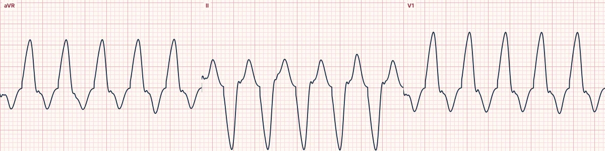
The idea
In SVT with aberrancy the first part of ventricular activation uses the normal, fast conduction system and moves away from aVR, so the QRS in aVR starts with a quick downward deflection. A VT from the apex or inferior wall moves toward aVR and writes an initial R wave, and any ventricular focus starts slowly.
vi/vt
Measure the vertical voltage change in the first 40 ms of the QRS (vi) and in the last 40 ms (vt). In aberrancy the start is fast and the delay comes late, so vi is larger. In VT the start is slow, so vi/vt is 1 or less.
Limits
Like other algorithms it performs less well with pre-excitation, drugs that slow conduction and very fast rates where the QRS onset and end are hard to see. Use it with, not instead of, AV dissociation and the clinical picture.
How to read it
- Find a clear QRS in aVR and mark its start and end.
- Ask the four questions in order and stop at the first yes.
- Confirm with AV dissociation, capture or fusion beats if you can find them.
Pitfalls
- Sodium-channel blocker toxicity gives a terminal, not initial, R in aVR: read which part of the QRS is positive.
- Measuring 40 ms at a fast rate needs a paused, magnified tracing.
Frequently asked questions
Vereckei aVR algorithm: what is the key idea?
A four-step method that diagnoses VT from lead aVR alone. It asks whether the start of the QRS in aVR is slow or points the wrong way, as happens when activation begins in the ventricle.
Vereckei aVR algorithm: what are the key numbers?
Lead: aVR only; Initial R in aVR: VT; Initial r or q: > 40 ms: VT; vi/vt: ≤ 1: VT.
Vereckei aVR algorithm: how do you apply it to an ECG?
Find a clear QRS in aVR and mark its start and end. Ask the four questions in order and stop at the first yes. Confirm with AV dissociation, capture or fusion beats if you can find them.
Vereckei aVR algorithm: what are the common pitfalls?
Sodium-channel blocker toxicity gives a terminal, not initial, R in aVR: read which part of the QRS is positive. Measuring 40 ms at a fast rate needs a paused, magnified tracing.