
Ventricular
Monomorphic ventricular tachycardia
A fast rhythm arising in the ventricles with every beat the same shape. It can be tolerated briefly, but often causes collapse and can degenerate into VF.
Last updated
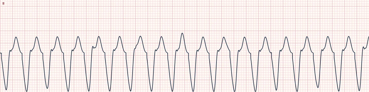
Example tracing, drawn by the lab's engine. Open the lab for a live, adjustable version.
At a glance
- Rate
- > 100/min, usually 120–250/min
- Rhythm
- Regular
- P waves
- Dissociated; often hidden
- QRS
- Wide (≥ 120 ms, often > 160 ms), uniform
Figures
Still images drawn by the lab's engine at 25 mm/s and 10 mm/mV, with the finding labeled. Select a figure to enlarge it.
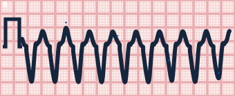
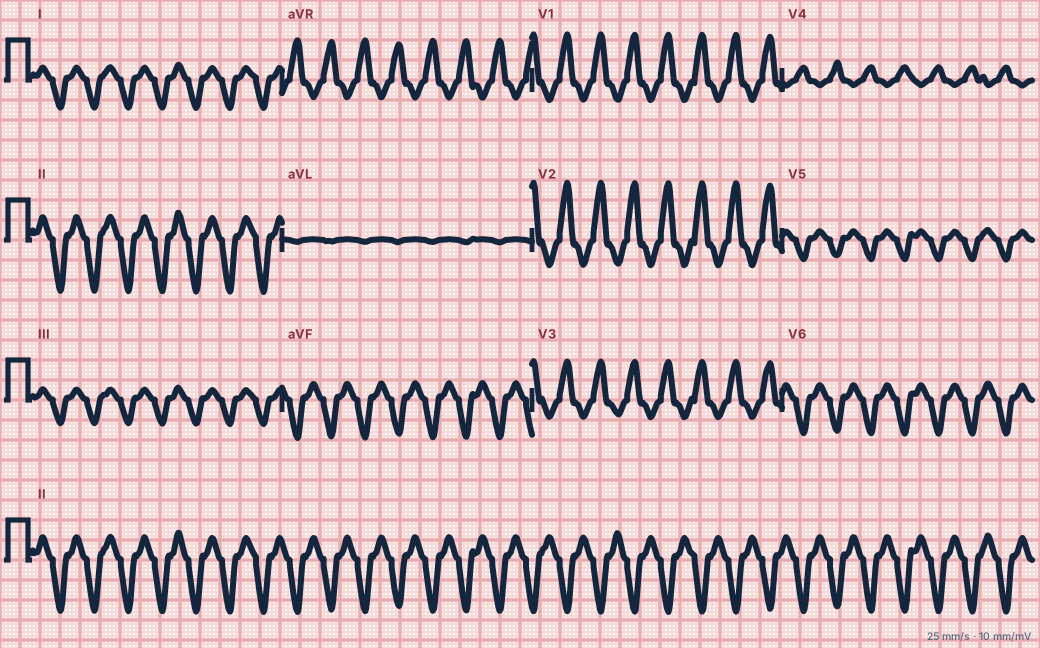
Scroll sideways to see the whole tracing, or select it to enlarge.
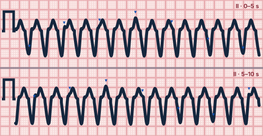
Scroll sideways to see the whole tracing, or select it to enlarge.
What the heart is doing
A re-entry circuit around ventricular scar, or a rapid focus, drives the ventricles from within the muscle. Activation bypasses the Purkinje network, and the atria beat on independently.
Why it looks this way
This is monomorphic VT because the tachycardia is regular with wide, uniform QRS complexes and atrial activity dissociated from the ventricles.
Why it matters
Check for a pulse. Pulseless VT is treated as cardiac arrest with CPR and defibrillation; unstable VT with a pulse needs immediate synchronized cardioversion; stable VT gets antiarrhythmics and expert help.
What to look for
- A regular, wide, fast rhythm: assume VT until proven otherwise.
- Look for P waves marching through at a slower rate, deforming occasional ST–T segments.
- Capture and fusion beats, or all chest leads pointing the same way, confirm VT.
Criteria
- Rate
- > 100/min, usually 120–250/min
- Rhythm
- Regular
- P waves
- Dissociated; often hidden
- QRS
- Wide (≥ 120 ms, often > 160 ms), uniform
- Axis
- Often extreme (negative in I and aVF)
Causes
- Scar from prior myocardial infarction
- Acute ischemia
- Cardiomyopathy and heart failure
- Hypokalemia, hypomagnesemia
- Drug toxicity
- Inherited arrhythmia syndromes
Can be mistaken for
- SVT with bundle branch block — typical RBBB or LBBB shape and a P wave tied to each QRS
- Antidromic AVRT — slurred delta-like onset in a patient with WPW Compare
- Hyperkalemia or sodium-channel blocker toxicity — very wide QRS at a slower rate
Advanced Down to the ion channel
What the cells are doing
Pacemaker, atrial and ventricular action potentials, in step with the tracing above, with sodium, calcium and potassium moving through their channels.
Frequently asked questions
What are the ECG criteria for monomorphic ventricular tachycardia?
Rate: > 100/min, usually 120–250/min. Rhythm: regular. P waves: dissociated; often hidden. QRS: wide (≥ 120 ms, often > 160 ms), uniform. Axis: often extreme (negative in I and aVF).
How is monomorphic ventricular tachycardia identified on an ECG?
This is monomorphic VT because the tachycardia is regular with wide, uniform QRS complexes and atrial activity dissociated from the ventricles.
What can monomorphic ventricular tachycardia be mistaken for?
SVT with bundle branch block: typical RBBB or LBBB shape and a P wave tied to each QRS. Antidromic AVRT: slurred delta-like onset in a patient with WPW. Hyperkalemia or sodium-channel blocker toxicity: very wide QRS at a slower rate.
What causes monomorphic ventricular tachycardia?
Common causes: scar from prior myocardial infarction; acute ischemia; cardiomyopathy and heart failure; hypokalemia, hypomagnesemia; drug toxicity; and inherited arrhythmia syndromes.
Practise it in the ACLS Lab
Sources
This page does not list page-specific sources yet.
Every source the site cites, with links