
Ventricular
Draft — pending owner review
Ventricular tachycardia with capture and fusion beats
Ventricular tachycardia while the sinus node keeps firing on its own. Now and then a P wave gets through: a narrow capture beat, or a hybrid fusion (Dressler) beat. Either proves the rhythm is VT.
Last updated
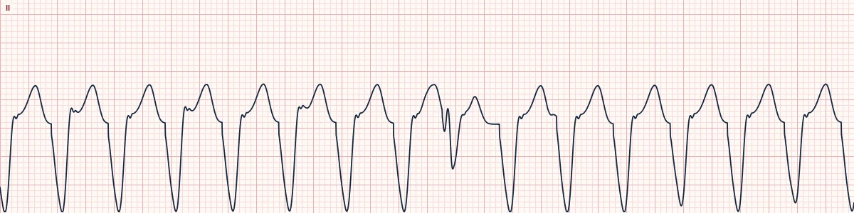
Example tracing, drawn by the lab's engine. Open the lab for a live, adjustable version.
At a glance
- Rate
- 120–180/min, regular apart from the captured beats
- QRS
- Wide (≥ 120 ms) in the VT beats
- AV relationship
- Dissociated: P waves march through at their own slower rate
- Capture beat
- An early, narrow QRS like the patient's normal beat
Figures
Still images drawn by the lab's engine at 25 mm/s and 10 mm/mV, with the finding labeled. Select a figure to enlarge it.
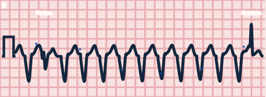
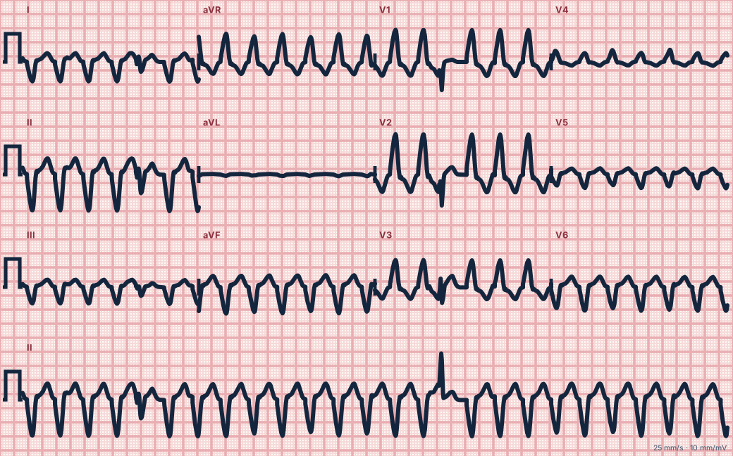
Scroll sideways to see the whole tracing, or select it to enlarge.
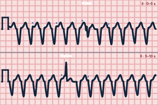
Scroll sideways to see the whole tracing, or select it to enlarge.
What the heart is doing
The ventricular focus and the sinus node beat independently. A sinus impulse that finds the AV node and ventricles recovered conducts normally (capture); one that arrives as the focus fires shares the ventricles with it (fusion).
Why it looks this way
This is VT with capture and fusion beats because wide regular complexes are dissociated from the P waves and are interrupted by narrow or intermediate beats.
Why it matters
Capture and fusion beats prove a ventricular origin, so the rhythm is treated as VT whatever the blood pressure. They are commonest when the VT is relatively slow, which leaves time for a sinus impulse to reach recovered ventricles.
What to look for
- March out the P waves: they keep their own rhythm straight through the wide complexes.
- Look for one early, narrow beat in a run of wide ones: a capture beat.
- A beat narrower than the rest but not normal in shape is a fusion beat.
Criteria
- Rate
- 120–180/min, regular apart from the captured beats
- QRS
- Wide (≥ 120 ms) in the VT beats
- AV relationship
- Dissociated: P waves march through at their own slower rate
- Capture beat
- An early, narrow QRS like the patient's normal beat
- Fusion beat
- A QRS part way between the VT beat and a normal beat
Causes
- Scar from a prior myocardial infarction
- Cardiomyopathy
- Acute ischemia
- Electrolyte disturbance
- Drug toxicity
Can be mistaken for
- SVT with aberrancy — P waves linked to every QRS, no fusion beats
- Accelerated idioventricular rhythm — rate under about 110/min Compare
- PVC — isolated wide beats in an otherwise sinus rhythm Compare
Advanced Down to the ion channel
What the cells are doing
Pacemaker, atrial and ventricular action potentials, in step with the tracing above, with sodium, calcium and potassium moving through their channels.
Frequently asked questions
What are the ECG criteria for ventricular tachycardia with capture and fusion beats?
Rate: 120–180/min, regular apart from the captured beats. QRS: wide (≥ 120 ms) in the VT beats. AV relationship: dissociated: P waves march through at their own slower rate. Capture beat: an early, narrow QRS like the patient's normal beat. Fusion beat: A QRS part way between the VT beat and a normal beat.
How is ventricular tachycardia with capture and fusion beats identified on an ECG?
This is VT with capture and fusion beats because wide regular complexes are dissociated from the P waves and are interrupted by narrow or intermediate beats.
What can ventricular tachycardia with capture and fusion beats be mistaken for?
SVT with aberrancy: P waves linked to every QRS, no fusion beats. Accelerated idioventricular rhythm: rate under about 110/min. PVC: isolated wide beats in an otherwise sinus rhythm.
What causes ventricular tachycardia with capture and fusion beats?
Common causes: scar from a prior myocardial infarction; cardiomyopathy; acute ischemia; electrolyte disturbance; and drug toxicity.
Sources
This page does not list page-specific sources yet.
Every source the site cites, with links