
Atrial
Atrial flutter, 4:1 conduction
A circuit spinning in the right atrium at about 300/min makes sawtooth flutter waves. The AV node lets every fourth one through, giving a ventricular rate near 75/min.
Last updated
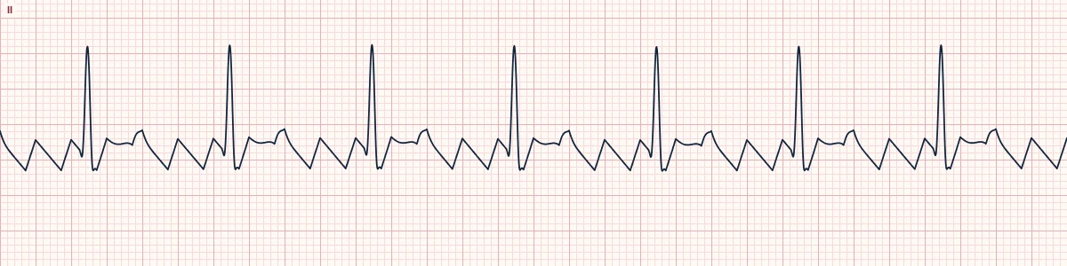
Example tracing, drawn by the lab's engine. Open the lab for a live, adjustable version.
At a glance
- Atrial rate
- 250–350/min (typically 300)
- Ventricular rate
- About 75/min, regular
- Flutter waves
- Sawtooth in II, III, aVF; small upright in V1
- Conduction
- 4 flutter waves for each QRS
Figures
Still images drawn by the lab's engine at 25 mm/s and 10 mm/mV, with the finding labeled. Select a figure to enlarge it.
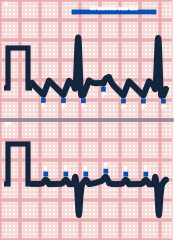
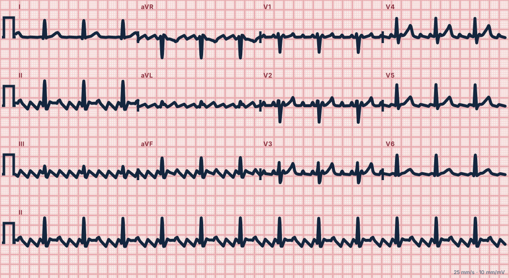
Scroll sideways to see the whole tracing, or select it to enlarge.
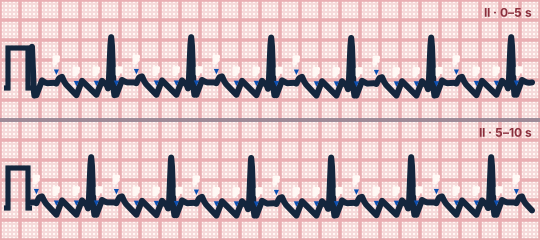
Scroll sideways to see the whole tracing, or select it to enlarge.
What the heart is doing
A re-entrant wavefront circles the right atrium around the tricuspid valve about 300 times a minute. The AV node cannot recover fast enough and conducts only every fourth circuit.
Why it looks this way
This is atrial flutter with 4:1 conduction because a sawtooth baseline at about 300/min has four flutter waves for each regular narrow QRS.
Why it matters
At this rate it is usually well tolerated, but the stroke risk is similar to atrial fibrillation and conduction can suddenly become 2:1. Management is rate or rhythm control plus stroke-risk assessment.
What to look for
- Look at II, III and aVF: the baseline never goes flat but slopes down and snaps up.
- Count the flutter waves per QRS, including the ones partly hidden by the QRS and T wave.
- V1 shows discrete upright atrial waves at about 300/min.
Criteria
- Atrial rate
- 250–350/min (typically 300)
- Ventricular rate
- About 75/min, regular
- Flutter waves
- Sawtooth in II, III, aVF; small upright in V1
- Conduction
- 4 flutter waves for each QRS
- QRS
- Narrow (< 120 ms)
Causes
- Right atrial enlargement, heart failure
- COPD and pulmonary hypertension
- Valvular and ischemic heart disease
- After cardiac surgery
- Hypertension
- Hyperthyroidism
Can be mistaken for
- Sinus rhythm with tremor or artifact — P waves can still be marched out
- Coarse atrial fibrillation — waves vary in shape and the R–R is irregular
- Atrial tachycardia with block — flat baseline between P waves
Advanced Down to the ion channel
What the cells are doing
Pacemaker, atrial and ventricular action potentials, in step with the tracing above, with sodium, calcium and potassium moving through their channels.
Frequently asked questions
What are the ECG criteria for atrial flutter, 4:1 conduction?
Atrial rate: 250–350/min (typically 300). Ventricular rate: about 75/min, regular. Flutter waves: sawtooth in II, III, aVF; small upright in V1. Conduction: 4 flutter waves for each QRS. QRS: narrow (< 120 ms).
How is atrial flutter, 4:1 conduction identified on an ECG?
This is atrial flutter with 4:1 conduction because a sawtooth baseline at about 300/min has four flutter waves for each regular narrow QRS.
What can atrial flutter, 4:1 conduction be mistaken for?
Sinus rhythm with tremor or artifact: P waves can still be marched out. Coarse atrial fibrillation: waves vary in shape and the R–R is irregular. Atrial tachycardia with block: flat baseline between P waves.
What causes atrial flutter, 4:1 conduction?
Common causes: right atrial enlargement, heart failure; COPD and pulmonary hypertension; valvular and ischemic heart disease; after cardiac surgery; hypertension; and hyperthyroidism.
Sources
This page does not list page-specific sources yet.
Every source the site cites, with links