
Atrial
Atrial flutter, 2:1 conduction
Atrial flutter in which every second flutter wave conducts, giving a regular narrow tachycardia at about 150/min. Two flutter waves belong to each QRS: one just before it and one in the ST segment.
Last updated
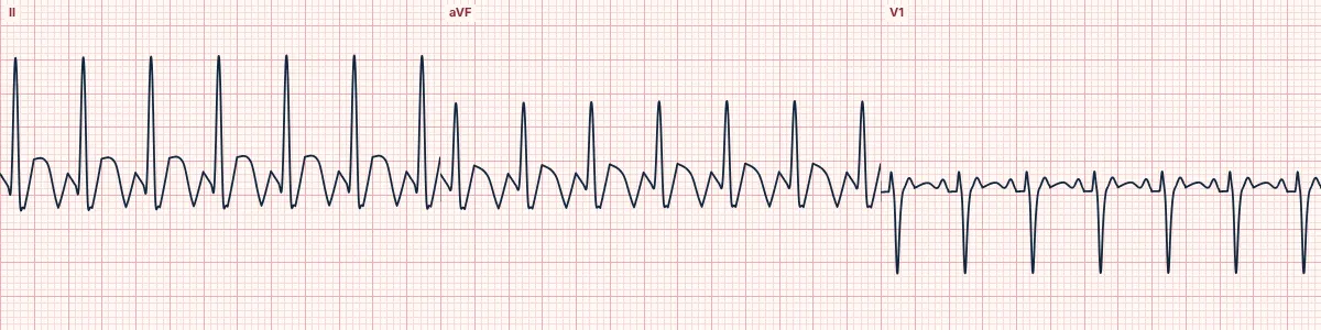
Example tracing, drawn by the lab's engine. Open the lab for a live, adjustable version.
At a glance
- Atrial rate
- 250–350/min (typically 300)
- Ventricular rate
- About 150/min, regular
- Flutter waves
- Sawtooth in II, III, aVF; one before each QRS and one in the ST segment
- Conduction
- 2 flutter waves for each QRS
Figures
Still images drawn by the lab's engine at 25 mm/s and 10 mm/mV, with the finding labeled. Select a figure to enlarge it.
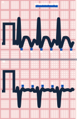
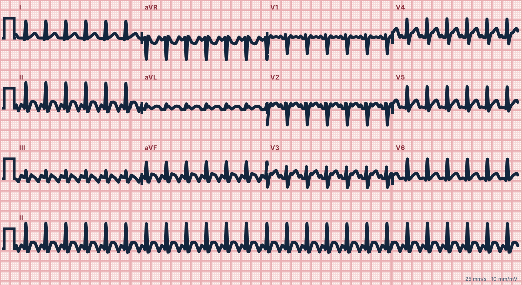
Scroll sideways to see the whole tracing, or select it to enlarge.
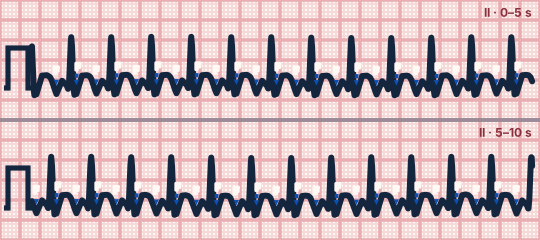
Scroll sideways to see the whole tracing, or select it to enlarge.
What the heart is doing
A right atrial re-entrant circuit runs at about 300/min. The AV node is refractory after each conducted impulse and blocks the next, so the ventricles follow at exactly half the atrial rate.
Why it looks this way
This is atrial flutter with 2:1 conduction because the regular narrow rhythm sits at about 150/min with two sawtooth waves for every QRS.
Why it matters
Unstable patients need immediate synchronized cardioversion. Stable patients get rate control and stroke-risk assessment. The rate barely changes with activity, a useful clue against sinus tachycardia.
What to look for
- Treat any regular narrow tachycardia fixed at about 150/min as flutter until proven otherwise.
- Find the two flutter waves for each QRS: one sits just before the QRS, the other in the ST segment, halfway between them.
- Look in II, aVF (a negative sawtooth) and V1 (small upright waves); a vagal maneuver or adenosine briefly unmasks the sawtooth.
Criteria
- Atrial rate
- 250–350/min (typically 300)
- Ventricular rate
- About 150/min, regular
- Flutter waves
- Sawtooth in II, III, aVF; one before each QRS and one in the ST segment
- Conduction
- 2 flutter waves for each QRS
- QRS
- Narrow (< 120 ms)
Causes
- Heart failure and atrial enlargement
- COPD, pulmonary embolism
- Valvular and ischemic heart disease
- After cardiac surgery
- Hyperthyroidism
- Alcohol
Can be mistaken for
- Sinus tachycardia — rate varies over time and there is one upright P per QRS Compare
- AVNRT — no atrial waves visible between the QRS complexes Compare
- Focal atrial tachycardia — flat baseline between P waves Compare
Advanced Down to the ion channel
What the cells are doing
Pacemaker, atrial and ventricular action potentials, in step with the tracing above, with sodium, calcium and potassium moving through their channels.
Frequently asked questions
What are the ECG criteria for atrial flutter, 2:1 conduction?
Atrial rate: 250–350/min (typically 300). Ventricular rate: about 150/min, regular. Flutter waves: sawtooth in II, III, aVF; one before each QRS and one in the ST segment. Conduction: 2 flutter waves for each QRS. QRS: narrow (< 120 ms).
How is atrial flutter, 2:1 conduction identified on an ECG?
This is atrial flutter with 2:1 conduction because the regular narrow rhythm sits at about 150/min with two sawtooth waves for every QRS.
What can atrial flutter, 2:1 conduction be mistaken for?
Sinus tachycardia: rate varies over time and there is one upright P per QRS. AVNRT: no atrial waves visible between the QRS complexes. Focal atrial tachycardia: flat baseline between P waves.
What causes atrial flutter, 2:1 conduction?
Common causes: heart failure and atrial enlargement; COPD, pulmonary embolism; valvular and ischemic heart disease; after cardiac surgery; hyperthyroidism; and alcohol.
Practise it in the ACLS Lab
Sources
This page does not list page-specific sources yet.
Every source the site cites, with links