
Atrial
Ectopic atrial rhythm
A steady rhythm at a normal rate driven by an atrial focus other than the sinus node, recognized by an abnormal P-wave shape or axis.
Last updated
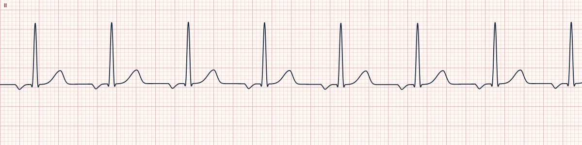
Example tracing, drawn by the lab's engine. Open the lab for a live, adjustable version.
At a glance
- Rate
- < 100/min
- Rhythm
- Regular
- P waves
- Non-sinus: here inverted in II, III and aVF
- PR interval
- ≥ 120 ms, constant
Figures
Still images drawn by the lab's engine at 25 mm/s and 10 mm/mV, with the finding labeled. Select a figure to enlarge it.
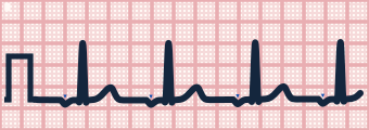
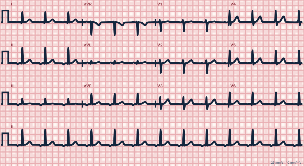
Scroll sideways to see the whole tracing, or select it to enlarge.
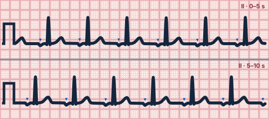
Scroll sideways to see the whole tracing, or select it to enlarge.
What the heart is doing
A focus low in the atrium fires slightly faster than the sinus node and takes over. The atria depolarize from bottom to top, inverting the P wave in the inferior leads; AV conduction is normal.
Why it looks this way
This is an ectopic atrial rhythm because inverted P waves with a PR of at least 120 ms precede every narrow QRS at a rate under 100/min.
Why it matters
Usually an incidental, harmless finding that needs no treatment. It matters mainly because it can be confused with lead misplacement or a junctional rhythm.
What to look for
- Check lead II first: a negative P wave before every QRS means the atria are being activated from below.
- Measure the PR. At 120 ms or more the focus is atrial; shorter than that suggests a junctional origin.
- Rule out misplaced limb electrodes before accepting an abnormal P axis.
Criteria
- Rate
- < 100/min
- Rhythm
- Regular
- P waves
- Non-sinus: here inverted in II, III and aVF
- PR interval
- ≥ 120 ms, constant
- QRS
- Narrow (< 120 ms)
Causes
- Normal variant, especially in the young and during sleep
- High vagal tone slowing the sinus node
- Sinus node dysfunction
- Atrial enlargement or scarring
- Digoxin effect
Can be mistaken for
- Accelerated junctional rhythm — retrograde P waves before the QRS (PR < 120 ms), within it or after it Compare
- Arm lead reversal — P, QRS and T are all inverted in lead I, not II Compare
- Normal sinus rhythm — P waves upright in II Compare
Advanced Down to the ion channel
What the cells are doing
Pacemaker, atrial and ventricular action potentials, in step with the tracing above, with sodium, calcium and potassium moving through their channels.
Frequently asked questions
What are the ECG criteria for ectopic atrial rhythm?
Rate: < 100/min. Rhythm: regular. P waves: non-sinus: here inverted in II, III and aVF. PR interval: ≥ 120 ms, constant. QRS: narrow (< 120 ms).
How is ectopic atrial rhythm identified on an ECG?
This is an ectopic atrial rhythm because inverted P waves with a PR of at least 120 ms precede every narrow QRS at a rate under 100/min.
What can ectopic atrial rhythm be mistaken for?
Accelerated junctional rhythm: retrograde P waves before the QRS (PR < 120 ms), within it or after it. Arm lead reversal: P, QRS and T are all inverted in lead I, not II. Normal sinus rhythm: P waves upright in II.
What causes ectopic atrial rhythm?
Common causes: normal variant, especially in the young and during sleep; high vagal tone slowing the sinus node; sinus node dysfunction; atrial enlargement or scarring; and digoxin effect.
Sources
This page does not list page-specific sources yet.
Every source the site cites, with links