
Atrial
Wandering atrial pacemaker
The pacemaker site shifts between the sinus node and other atrial sites, so P waves keep changing shape while the rate stays under 100/min.
Last updated
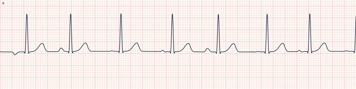
Example tracing, drawn by the lab's engine. Open the lab for a live, adjustable version.
At a glance
- Rate
- < 100/min
- Rhythm
- Irregularly irregular
- P waves
- At least 3 different shapes in one lead
- PR interval
- Varies from beat to beat
Figures
Still images drawn by the lab's engine at 25 mm/s and 10 mm/mV, with the finding labeled. Select a figure to enlarge it.
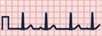
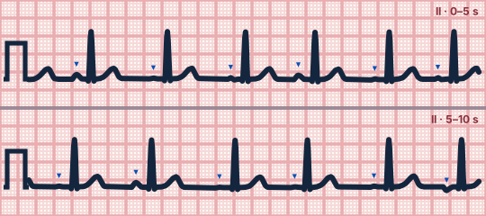
Scroll sideways to see the whole tracing, or select it to enlarge.
What the heart is doing
Several atrial sites take turns as pacemaker. Each site activates the atria by a different route and reaches the AV node after a different delay, changing the P-wave shape and PR.
Why it looks this way
This is a wandering atrial pacemaker because there are at least three P-wave shapes with varying PR intervals at a rate below 100/min.
Why it matters
Generally benign and rarely needs treatment. The same pattern above 100/min is multifocal atrial tachycardia, which signals significant underlying illness.
What to look for
- Compare P waves along one lead: you should find three or more shapes (upright, flat, inverted).
- There is a P wave before every QRS, which separates this from atrial fibrillation.
- The baseline between P waves is flat, with no fibrillatory or flutter waves.
Criteria
- Rate
- < 100/min
- Rhythm
- Irregularly irregular
- P waves
- At least 3 different shapes in one lead
- PR interval
- Varies from beat to beat
- QRS
- Narrow (< 120 ms)
Causes
- High vagal tone in the young and athletic
- Sleep
- Chronic lung disease
- Digoxin effect
- Sinus node dysfunction
Can be mistaken for
- Atrial fibrillation — no distinct P waves at all Compare
- Multifocal atrial tachycardia — the same pattern at a rate above 100/min Compare
- Sinus rhythm with frequent PACs — one dominant P wave with occasional early beats
Advanced Down to the ion channel
What the cells are doing
Pacemaker, atrial and ventricular action potentials, in step with the tracing above, with sodium, calcium and potassium moving through their channels.
Frequently asked questions
What are the ECG criteria for wandering atrial pacemaker?
Rate: < 100/min. Rhythm: irregularly irregular. P waves: at least 3 different shapes in one lead. PR interval: varies from beat to beat. QRS: narrow (< 120 ms).
How is wandering atrial pacemaker identified on an ECG?
This is a wandering atrial pacemaker because there are at least three P-wave shapes with varying PR intervals at a rate below 100/min.
What can wandering atrial pacemaker be mistaken for?
Atrial fibrillation: no distinct P waves at all. Multifocal atrial tachycardia: the same pattern at a rate above 100/min. Sinus rhythm with frequent PACs: one dominant P wave with occasional early beats.
What causes wandering atrial pacemaker?
Common causes: high vagal tone in the young and athletic; sleep; chronic lung disease; digoxin effect; and sinus node dysfunction.
Sources
This page does not list page-specific sources yet.
Every source the site cites, with links