
Other conditions
Pulmonary embolism (right heart strain)
A clot in the pulmonary arteries abruptly loads the right ventricle. The ECG may show right heart strain, though it is often just a fast sinus rhythm.
Last updated
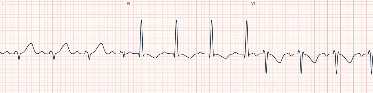
Example tracing, drawn by the lab's engine. Open the lab for a live, adjustable version.
At a glance
- Rate
- Sinus tachycardia is the most common finding
- S1Q3T3
- Deep S in I, Q wave and inverted T in III
- T waves
- Inverted in the right chest leads (V1–V3, sometimes V4) and in III
- QRS
- New incomplete or complete RBBB
Figures
Still images drawn by the lab's engine at 25 mm/s and 10 mm/mV, with the finding labeled. Select a figure to enlarge it.
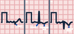
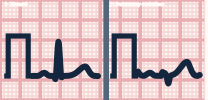
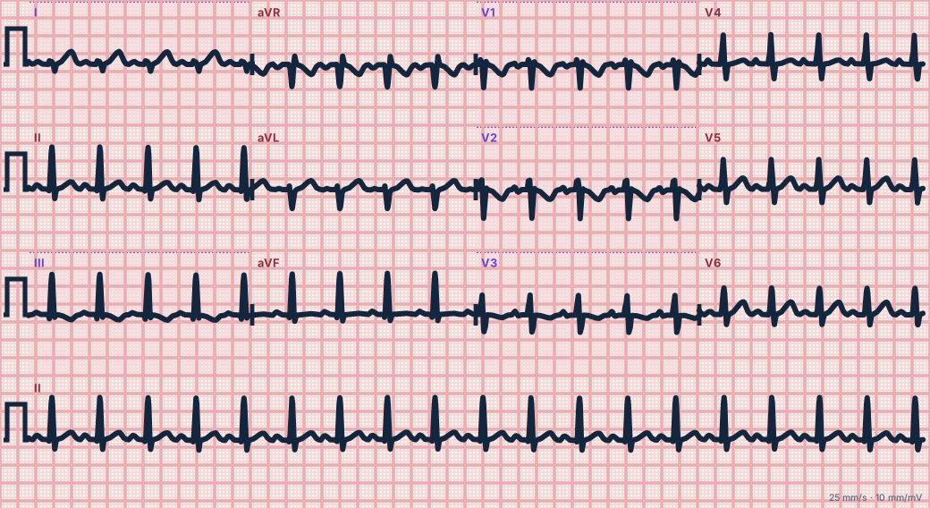
Scroll sideways to see the whole tracing, or select it to enlarge.
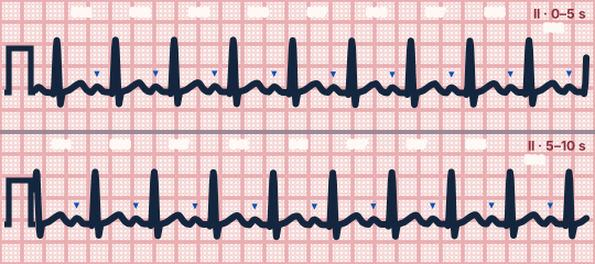
Scroll sideways to see the whole tracing, or select it to enlarge.
What the heart is doing
Sudden obstruction of the pulmonary arteries raises right ventricular pressure. The right ventricle dilates and becomes ischemic, delaying its activation and reversing its repolarization.
Why it looks this way
This is pulmonary embolism with right heart strain because an S1Q3T3 pattern accompanies T-wave inversion in the right chest leads and a rightward axis shift.
Why it matters
The ECG cannot rule PE in or out, but signs of right heart strain point to a larger clot burden. Shock or collapse indicates a massive embolism needing immediate reperfusion treatment.
What to look for
- T-wave inversion in both the right chest leads and lead III strongly favors PE over ischemia.
- A new S wave in lead I with a new Q wave in III.
- Unexplained tachycardia and breathlessness matter more than any ECG sign.
Criteria
- Rate
- Sinus tachycardia is the most common finding
- S1Q3T3
- Deep S in I, Q wave and inverted T in III
- T waves
- Inverted in the right chest leads (V1–V3, sometimes V4) and in III
- QRS
- New incomplete or complete RBBB
- Axis
- Shifted rightward
Causes
- Deep vein thrombosis
- Recent surgery or immobilization
- Cancer
- Pregnancy and estrogen therapy
- Previous clots or inherited clotting disorders
- Long-distance travel
Can be mistaken for
- Wellens pattern — T inversion in V2–V3 without inversion in lead III
- Old inferior infarction — Q waves in II and aVF as well as III Compare
- Chronic lung disease — long-standing right axis and P pulmonale
Advanced Down to the ion channel
What the cells are doing
Pacemaker, atrial and ventricular action potentials, in step with the tracing above, with sodium, calcium and potassium moving through their channels.
Frequently asked questions
What are the ECG criteria for pulmonary embolism (right heart strain)?
Rate: sinus tachycardia is the most common finding. S1Q3T3: deep S in I, Q wave and inverted T in III. T waves: inverted in the right chest leads (V1–V3, sometimes V4) and in III. QRS: new incomplete or complete RBBB. Axis: shifted rightward.
How is pulmonary embolism (right heart strain) identified on an ECG?
This is pulmonary embolism with right heart strain because an S1Q3T3 pattern accompanies T-wave inversion in the right chest leads and a rightward axis shift.
What can pulmonary embolism (right heart strain) be mistaken for?
Wellens pattern: T inversion in V2–V3 without inversion in lead III. Old inferior infarction: Q waves in II and aVF as well as III. Chronic lung disease: long-standing right axis and P pulmonale.
What causes pulmonary embolism (right heart strain)?
Common causes: deep vein thrombosis; recent surgery or immobilization; cancer; pregnancy and estrogen therapy; previous clots or inherited clotting disorders; and long-distance travel.
Practise it in the ACLS Lab
Sources
This page does not list page-specific sources yet.
Every source the site cites, with links