
Repolarization & channelopathies
Brugada pattern, type 1
An inherited sodium-channel disorder that shows as coved ST elevation in V1–V2. The heart is structurally normal but prone to ventricular fibrillation.
Last updated
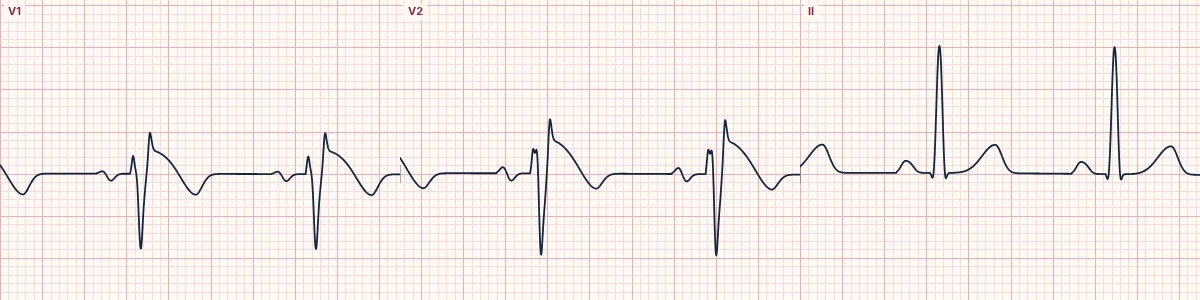
Example tracing, drawn by the lab's engine. Open the lab for a live, adjustable version.
At a glance
- V1–V2
- Coved ST elevation ≥ 2 mm, sloping down into an inverted T wave
- Leads
- At least one of V1 or V2
- QRS
- Pseudo-RBBB shape without a wide S wave in I or V6
- Other leads
- Normal
Figures
Still images drawn by the lab's engine at 25 mm/s and 10 mm/mV, with the finding labeled. Select a figure to enlarge it.
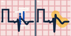
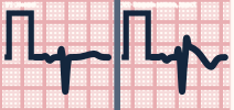
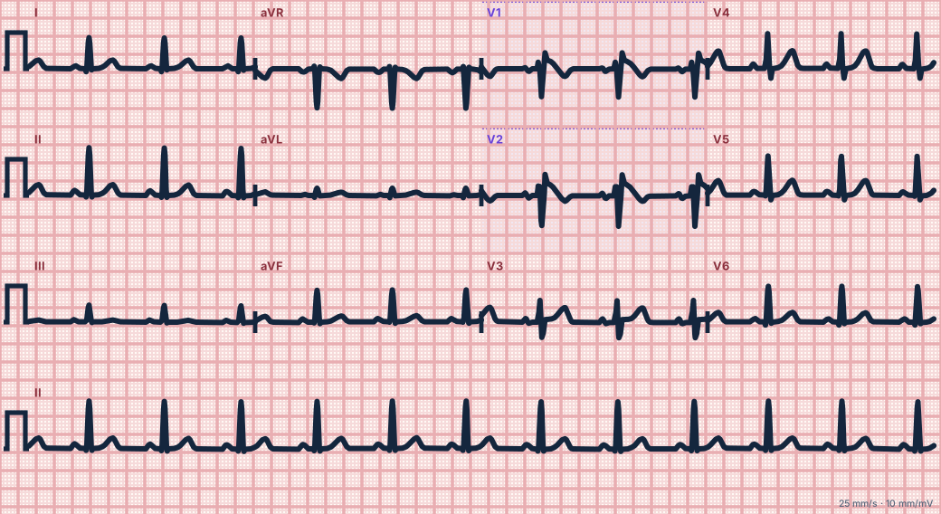
Scroll sideways to see the whole tracing, or select it to enlarge.
What the heart is doing
Reduced sodium current leaves outward currents unopposed in the right ventricular outflow tract's outer layer. The resulting voltage gradient across the wall raises the ST segment in V1–V2.
Why it looks this way
This is the Brugada type 1 pattern because V1–V2 show coved ST elevation of at least 2 mm descending into an inverted T wave.
Why it matters
With syncope, nocturnal gasping, family sudden death or documented arrhythmia it is Brugada syndrome, with a risk of sudden death. Treat fever, avoid provoking drugs and refer urgently.
What to look for
- In V1–V2, the ST rises high at the J point and descends in a convex curve to a negative T wave.
- Check I and V6: without a wide terminal S wave, this is not a true RBBB.
- Moving V1–V2 up one or two rib spaces can unmask the pattern.
Criteria
- V1–V2
- Coved ST elevation ≥ 2 mm, sloping down into an inverted T wave
- Leads
- At least one of V1 or V2
- QRS
- Pseudo-RBBB shape without a wide S wave in I or V6
- Other leads
- Normal
Causes
- Inherited sodium-channel variants
- Unmasked by fever
- Unmasked by sodium-channel blocking drugs
- Unmasked by alcohol or cocaine
- More common in men and in Southeast Asian populations
Can be mistaken for
- RBBB — rsR′ in V1 with a wide S in I and V6 and no coved elevation Compare
- Anteroseptal STEMI — chest pain, elevation extending to V3–V4, reciprocal changes
- Early repolarization — concave ST elevation with an upright T wave Compare
Advanced Down to the ion channel
What the cells are doing
Pacemaker, atrial and ventricular action potentials, in step with the tracing above, with sodium, calcium and potassium moving through their channels.
Frequently asked questions
What are the ECG criteria for brugada pattern, type 1?
V1–V2: coved ST elevation ≥ 2 mm, sloping down into an inverted T wave. Leads: at least one of V1 or V2. QRS: pseudo-RBBB shape without a wide S wave in I or V6. Other leads: normal.
How is brugada pattern, type 1 identified on an ECG?
This is the Brugada type 1 pattern because V1–V2 show coved ST elevation of at least 2 mm descending into an inverted T wave.
What can brugada pattern, type 1 be mistaken for?
RBBB: rsR′ in V1 with a wide S in I and V6 and no coved elevation. Anteroseptal STEMI: chest pain, elevation extending to V3–V4, reciprocal changes. Early repolarization: concave ST elevation with an upright T wave.
What causes brugada pattern, type 1?
Common causes: inherited sodium-channel variants; unmasked by fever; unmasked by sodium-channel blocking drugs; unmasked by alcohol or cocaine; and more common in men and in Southeast Asian populations.
Sources
This page does not list page-specific sources yet.
Every source the site cites, with links