
Repolarization & channelopathies
Long QT
Ventricular repolarization takes too long, shown by a prolonged QT interval. Inherited or acquired, it predisposes to torsades de pointes.
Last updated
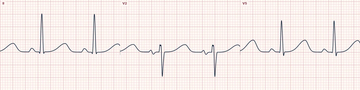
Example tracing, drawn by the lab's engine. Open the lab for a live, adjustable version.
At a glance
- QTc
- > 450 ms in men, > 460 ms in women
- High risk
- QTc > 500 ms
- T waves
- Broad, late, sometimes notched
- QRS
- Normal
Figures
Still images drawn by the lab's engine at 25 mm/s and 10 mm/mV, with the finding labeled. Select a figure to enlarge it.
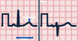
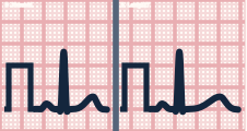
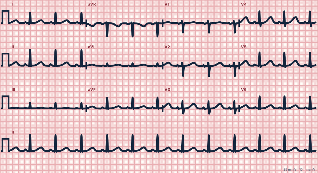
Scroll sideways to see the whole tracing, or select it to enlarge.
What the heart is doing
Reduced outward potassium current or persistent inward sodium current lengthens the action potential. Prolonged, uneven repolarization lets early afterdepolarizations trigger extra beats.
Why it looks this way
This is long QT because the rate-corrected QT interval is well above 460 ms, with the T wave ending past the midpoint of the R–R interval.
Why it matters
The longer the QT, the higher the risk of torsades de pointes, syncope and sudden death. Stop offending drugs, correct electrolytes and avoid adding other QT-prolonging medications.
What to look for
- Quick check at normal rates: the QT should be less than half the R–R interval.
- Measure in II or V5 to the end of the T wave, and correct for rate.
- Do not include a separate U wave in the measurement.
Criteria
- QTc
- > 450 ms in men, > 460 ms in women
- High risk
- QTc > 500 ms
- T waves
- Broad, late, sometimes notched
- QRS
- Normal
Causes
- QT-prolonging drugs (antiarrhythmics, antipsychotics, some antibiotics, methadone)
- Hypokalemia, hypomagnesemia, hypocalcemia
- Congenital ion channel disorders
- Bradycardia and heart block
- Hypothermia
- Brain injury
Can be mistaken for
- Hypokalemia — a T–U complex mistaken for a long QT Compare
- Hypocalcemia — the ST segment is long while the T wave is normal Compare
- Bundle branch block — the QT is long only because the QRS is wide
Advanced Down to the ion channel
What the cells are doing
Pacemaker, atrial and ventricular action potentials, in step with the tracing above, with sodium, calcium and potassium moving through their channels.
Frequently asked questions
What are the ECG criteria for long QT?
QTc: > 450 ms in men, > 460 ms in women. High risk: QTc > 500 ms. T waves: broad, late, sometimes notched. QRS: normal.
How is long QT identified on an ECG?
This is long QT because the rate-corrected QT interval is well above 460 ms, with the T wave ending past the midpoint of the R–R interval.
What can long QT be mistaken for?
Hypokalemia: a T–U complex mistaken for a long QT. Hypocalcemia: the ST segment is long while the T wave is normal. Bundle branch block: the QT is long only because the QRS is wide.
What causes long QT?
Common causes: QT-prolonging drugs (antiarrhythmics, antipsychotics, some antibiotics, methadone); hypokalemia, hypomagnesemia, hypocalcemia; congenital ion channel disorders; bradycardia and heart block; hypothermia; and brain injury.
Practise it in the ACLS Lab
Sources
This page does not list page-specific sources yet.
Every source the site cites, with links