
Repolarization & channelopathies
Short QT
Ventricular repolarization is abnormally fast, giving a very short QT with tall, peaked T waves. A rare inherited cause of sudden death.
Last updated
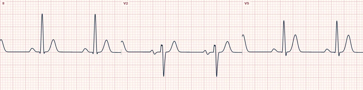
Example tracing, drawn by the lab's engine. Open the lab for a live, adjustable version.
At a glance
- QTc
- ≤ 320 ms; or ≤ 360 ms with a pathogenic mutation, family history of short QT or survived VT/VF arrest
- ST segment
- Absent or very short
- T waves
- Tall, narrow, symmetric, especially in the chest leads
- Rate response
- QT barely changes with heart rate
Figures
Still images drawn by the lab's engine at 25 mm/s and 10 mm/mV, with the finding labeled. Select a figure to enlarge it.
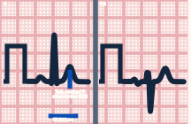
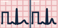
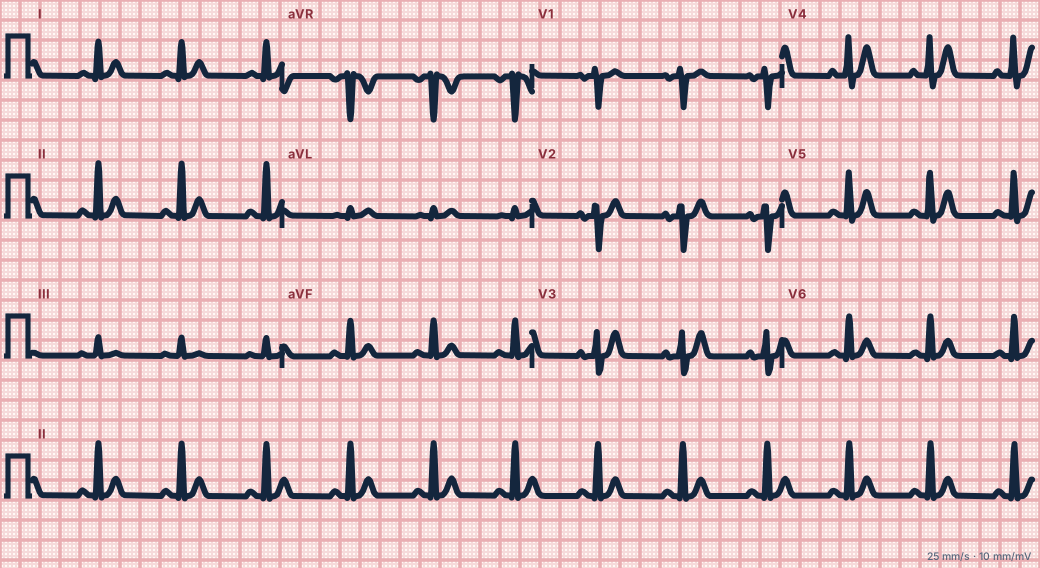
Scroll sideways to see the whole tracing, or select it to enlarge.
What the heart is doing
Overactive potassium channels repolarize the ventricles too quickly. The refractory period is short and uneven, making it easy for re-entry to start in both atria and ventricles.
Why it looks this way
This is short QT because the corrected QT interval is 320 ms or less, with tall peaked T waves and almost no ST segment.
Why it matters
The congenital form carries a risk of atrial fibrillation at a young age and of ventricular fibrillation. First exclude reversible causes, then refer for specialist assessment.
What to look for
- The T wave rises straight from the QRS with no ST segment.
- Tall, pointed T waves in a young patient without hyperkalemia.
- Ask about fainting, palpitations and sudden death in relatives.
Criteria
- QTc
- ≤ 320 ms; or ≤ 360 ms with a pathogenic mutation, family history of short QT or survived VT/VF arrest
- ST segment
- Absent or very short
- T waves
- Tall, narrow, symmetric, especially in the chest leads
- Rate response
- QT barely changes with heart rate
Causes
- Inherited potassium-channel gain of function
- Hypercalcemia
- Hyperkalemia
- Digoxin
- Acidosis
- Hyperthermia
Can be mistaken for
- Hypercalcemia — short ST but normal T-wave height Compare
- Hyperkalemia — peaked T waves with a normal or only slightly short QT
- Digoxin effect — short QT with scooped ST depression Compare
Advanced Down to the ion channel
What the cells are doing
Pacemaker, atrial and ventricular action potentials, in step with the tracing above, with sodium, calcium and potassium moving through their channels.
Frequently asked questions
What are the ECG criteria for short QT?
QTc: ≤ 320 ms; or ≤ 360 ms with a pathogenic mutation, family history of short QT or survived VT/VF arrest. ST segment: absent or very short. T waves: tall, narrow, symmetric, especially in the chest leads. Rate response: QT barely changes with heart rate.
How is short QT identified on an ECG?
This is short QT because the corrected QT interval is 320 ms or less, with tall peaked T waves and almost no ST segment.
What can short QT be mistaken for?
Hypercalcemia: short ST but normal T-wave height. Hyperkalemia: peaked T waves with a normal or only slightly short QT. Digoxin effect: short QT with scooped ST depression.
What causes short QT?
Common causes: inherited potassium-channel gain of function; hypercalcemia; hyperkalemia; digoxin; acidosis; and hyperthermia.
Sources
This page does not list page-specific sources yet.
Every source the site cites, with links