
Electrolytes
Hypocalcemia
Low blood calcium lengthens the plateau of the action potential. The ST segment is stretched out, prolonging the QT, while the T wave keeps its normal width.
Last updated
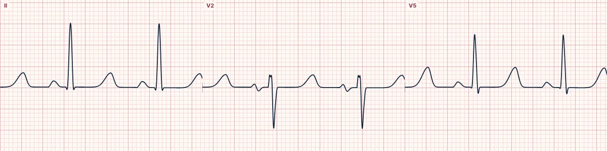
Example tracing, drawn by the lab's engine. Open the lab for a live, adjustable version.
At a glance
- QT
- Prolonged (QTc > 460 ms)
- ST segment
- Long and flat
- T waves
- Normal width, arriving late
- QRS
- Normal
Figures
Still images drawn by the lab's engine at 25 mm/s and 10 mm/mV, with the finding labeled. Select a figure to enlarge it.
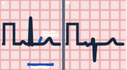
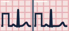
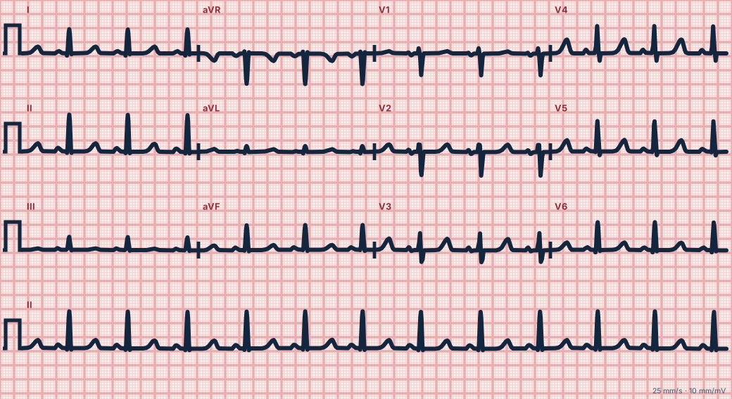
Scroll sideways to see the whole tracing, or select it to enlarge.
What the heart is doing
Low calcium prolongs phase 2 (the plateau) of the ventricular action potential. The plateau corresponds to the ST segment, so the ST lengthens while the T wave itself is unchanged.
Why it looks this way
This is hypocalcemia because the QT is prolonged by a long, flat ST segment while the T wave keeps a normal width.
Why it matters
Clinically causes tingling, muscle spasm, laryngospasm and seizures. Torsades de pointes is less common than with other causes of long QT but possible. Symptomatic patients need calcium replacement.
What to look for
- A long flat stretch between the end of the QRS and the start of the T wave.
- The T wave is a normal size and shape; it is simply late.
- No U waves, which separates it from hypokalemia.
Criteria
- QT
- Prolonged (QTc > 460 ms)
- ST segment
- Long and flat
- T waves
- Normal width, arriving late
- QRS
- Normal
Causes
- Hypoparathyroidism, including after neck surgery
- Chronic kidney disease
- Vitamin D deficiency
- Acute pancreatitis
- Massive blood transfusion (citrate)
- Hypomagnesemia
Can be mistaken for
- Drug-induced or congenital long QT — the T wave itself is broad or misshapen
- Hypokalemia — flat T waves with U waves Compare
- Hypothermia — long QT with J waves and bradycardia Compare
Advanced Down to the ion channel
What the cells are doing
Pacemaker, atrial and ventricular action potentials, in step with the tracing above, with sodium, calcium and potassium moving through their channels.
Frequently asked questions
What are the ECG criteria for hypocalcemia?
QT: prolonged (QTc > 460 ms). ST segment: long and flat. T waves: normal width, arriving late. QRS: normal.
How is hypocalcemia identified on an ECG?
This is hypocalcemia because the QT is prolonged by a long, flat ST segment while the T wave keeps a normal width.
What can hypocalcemia be mistaken for?
Drug-induced or congenital long QT: the T wave itself is broad or misshapen. Hypokalemia: flat T waves with U waves. Hypothermia: long QT with J waves and bradycardia.
What causes hypocalcemia?
Common causes: hypoparathyroidism, including after neck surgery; chronic kidney disease; vitamin D deficiency; acute pancreatitis; massive blood transfusion (citrate); and hypomagnesemia.
Sources
This page does not list page-specific sources yet.
Every source the site cites, with links