
Ischemia & prior infarction
de Winter T waves
An acute LAD occlusion that shows no ST elevation in the chest leads. The ST segment instead starts below baseline and climbs steeply into a tall, symmetric T wave.
Last updated
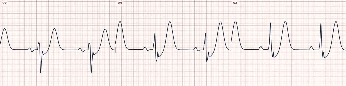
Example tracing, drawn by the lab's engine. Open the lab for a live, adjustable version.
At a glance
- ST segment
- Upsloping depression of 1–3 mm at the J point in V2–V6
- T waves
- Tall, symmetric, prominent in V2–V4
- aVR
- Often 0.5–1 mm of ST elevation
- QRS
- Normal or slightly wide; R waves may be reduced
Figures
Still images drawn by the lab's engine at 25 mm/s and 10 mm/mV, with the finding labeled. Select a figure to enlarge it.
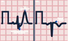
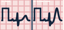
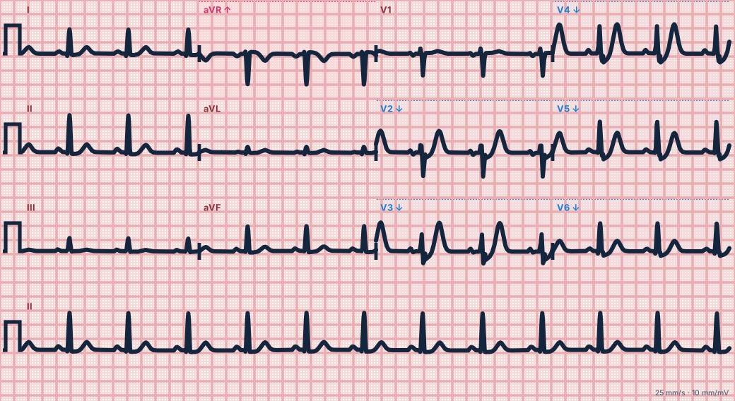
Scroll sideways to see the whole tracing, or select it to enlarge.
What the heart is doing
Acute anterior ischemia, possibly limited to the inner wall with delayed conduction, produces tall repolarization forces toward the chest leads while the ST takeoff is pulled below baseline.
Why it looks this way
This is the de Winter pattern because upsloping ST depression at the J point leads into tall symmetric T waves in the precordial leads.
Why it matters
A STEMI equivalent: the artery is occluded even though classic ST elevation is absent. Treat as an anterior STEMI and activate the cath lab without waiting for the ECG to evolve.
What to look for
- The J point sits below the baseline and the ST rises straight into a tall T wave.
- There is no ST elevation in the precordial leads despite ongoing chest pain.
- Look for slight ST elevation in aVR.
Criteria
- ST segment
- Upsloping depression of 1–3 mm at the J point in V2–V6
- T waves
- Tall, symmetric, prominent in V2–V4
- aVR
- Often 0.5–1 mm of ST elevation
- QRS
- Normal or slightly wide; R waves may be reduced
Causes
- Acute proximal LAD occlusion
- Plaque rupture with thrombosis
- Seen more in younger men with high cholesterol
- Coronary artery spasm (rare)
Can be mistaken for
- Hyperkalemia — narrow tented T waves in most leads without J-point depression
- Rate-related ST depression — upsloping, but the T waves are not tall
- Early repolarization — the J point is elevated, not depressed Compare
Advanced Down to the ion channel
What the cells are doing
Pacemaker, atrial and ventricular action potentials, in step with the tracing above, with sodium, calcium and potassium moving through their channels.
Frequently asked questions
What are the ECG criteria for de Winter T waves?
ST segment: upsloping depression of 1–3 mm at the J point in V2–V6. T waves: tall, symmetric, prominent in V2–V4. aVR: often 0.5–1 mm of ST elevation. QRS: normal or slightly wide; R waves may be reduced.
How is de Winter T waves identified on an ECG?
This is the de Winter pattern because upsloping ST depression at the J point leads into tall symmetric T waves in the precordial leads.
What can de Winter T waves be mistaken for?
Hyperkalemia: narrow tented T waves in most leads without J-point depression. Rate-related ST depression: upsloping, but the T waves are not tall. Early repolarization: the J point is elevated, not depressed.
What causes de Winter T waves?
Common causes: acute proximal LAD occlusion; plaque rupture with thrombosis; seen more in younger men with high cholesterol; and coronary artery spasm (rare).
Sources
This page does not list page-specific sources yet.
Every source the site cites, with links