
Ischemia & prior infarction
Wellens pattern, type A (biphasic T)
A warning sign of a critical narrowing in the proximal LAD, seen when the patient is pain-free: the T wave in V2–V3 goes up, then dips below the baseline.
Last updated
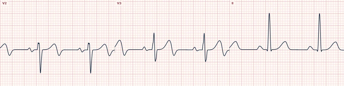
Example tracing, drawn by the lab's engine. Open the lab for a live, adjustable version.
At a glance
- T waves
- Biphasic (up then down) in V2–V3, sometimes V4
- ST segment
- Isoelectric or < 1 mm elevated
- QRS
- No Q waves; R-wave progression preserved
- Context
- Recent chest pain, now resolved
Figures
Still images drawn by the lab's engine at 25 mm/s and 10 mm/mV, with the finding labeled. Select a figure to enlarge it.
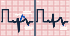
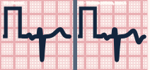
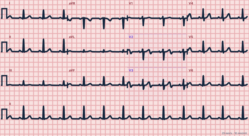
Scroll sideways to see the whole tracing, or select it to enlarge.
What the heart is doing
After a brief LAD occlusion reopens, the anterior wall repolarizes abnormally. The last part of repolarization reverses direction, producing a terminal negative deflection on the T wave.
Why it looks this way
This is Wellens pattern type A because the T waves in V2–V3 are biphasic, positive then negative, with no significant ST elevation or Q waves.
Why it matters
Signals a high risk of a large anterior infarction within days to weeks. Treat as an acute coronary syndrome needing urgent angiography; stress testing is dangerous and should be avoided.
What to look for
- Check the end of the T wave in V2 and V3: it finishes with a sharp downward dip.
- R waves are intact and there is no significant ST elevation.
- The pattern appears between episodes of pain and may normalize during pain.
Criteria
- T waves
- Biphasic (up then down) in V2–V3, sometimes V4
- ST segment
- Isoelectric or < 1 mm elevated
- QRS
- No Q waves; R-wave progression preserved
- Context
- Recent chest pain, now resolved
Causes
- Critical proximal LAD stenosis
- Spontaneous reperfusion of an LAD occlusion
- Coronary artery spasm
- Unstable plaque with intermittent occlusion
Can be mistaken for
- Normal variant T waves — the terminal part is not sharply negative
- Hypokalemia — a down-up T–U complex, the opposite sequence Compare
- Pulmonary embolism — T inversion in V1–V4 together with lead III Compare
Advanced Down to the ion channel
What the cells are doing
Pacemaker, atrial and ventricular action potentials, in step with the tracing above, with sodium, calcium and potassium moving through their channels.
Frequently asked questions
What are the ECG criteria for wellens pattern, type A (biphasic T)?
T waves: biphasic (up then down) in V2–V3, sometimes V4. ST segment: isoelectric or < 1 mm elevated. QRS: no Q waves; R-wave progression preserved. Context: recent chest pain, now resolved.
How is wellens pattern, type A (biphasic T) identified on an ECG?
This is Wellens pattern type A because the T waves in V2–V3 are biphasic, positive then negative, with no significant ST elevation or Q waves.
What can wellens pattern, type A (biphasic T) be mistaken for?
Normal variant T waves: the terminal part is not sharply negative. Hypokalemia: a down-up T–U complex, the opposite sequence. Pulmonary embolism: T inversion in V1–V4 together with lead III.
What causes wellens pattern, type A (biphasic T)?
Common causes: critical proximal LAD stenosis; spontaneous reperfusion of an LAD occlusion; coronary artery spasm; and unstable plaque with intermittent occlusion.
Sources
This page does not list page-specific sources yet.
Every source the site cites, with links