
Drugs & toxins
Digoxin effect
The expected ECG signature of a patient taking digoxin: scooped, sagging ST segments with a short QT. It shows the drug is present, not that the level is toxic.
Last updated
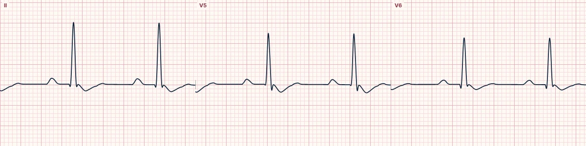
Example tracing, drawn by the lab's engine. Open the lab for a live, adjustable version.
At a glance
- ST segment
- Downsloping, scooped depression
- T waves
- Flattened, inverted or biphasic
- QT
- Shortened
- PR interval
- Mildly prolonged
Figures
Still images drawn by the lab's engine at 25 mm/s and 10 mm/mV, with the finding labeled. Select a figure to enlarge it.
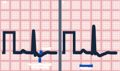
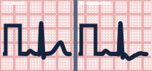
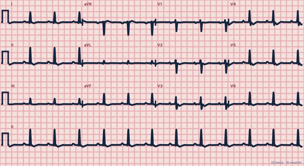
Scroll sideways to see the whole tracing, or select it to enlarge.
What the heart is doing
Digoxin inhibits the sodium–potassium pump, raising calcium inside cells and shortening the action potential. It also increases vagal tone, slowing AV nodal conduction.
Why it looks this way
This is digoxin effect because the ST segments are scooped and downsloping with flattened T waves and a short QT.
Why it matters
Not harmful in itself. Suspect toxicity when new arrhythmias appear: frequent PVCs, atrial tachycardia with block, junctional rhythms or AV block. Low potassium and kidney failure raise the risk.
What to look for
- ST segments that sag smoothly downward like a hockey stick or a reversed tick.
- A short QT, unlike the long QT of hypokalemia.
- Check the rhythm separately; toxicity shows as arrhythmias, not as deeper scooping.
Criteria
- ST segment
- Downsloping, scooped depression
- T waves
- Flattened, inverted or biphasic
- QT
- Shortened
- PR interval
- Mildly prolonged
- Leads
- Most obvious where the R wave is tall (lateral leads)
Causes
- Therapeutic digoxin use
- Rate control in atrial fibrillation
- Heart failure treatment
- Other cardiac glycosides (plant poisonings)
Can be mistaken for
- Ischemia — horizontal or downsloping ST depression with symptoms and a normal QT
- LVH with strain — high voltage and asymmetric T inversion Compare
- Hypokalemia — ST depression with U waves and a long QT Compare
Advanced Down to the ion channel
What the cells are doing
Pacemaker, atrial and ventricular action potentials, in step with the tracing above, with sodium, calcium and potassium moving through their channels.
Frequently asked questions
What are the ECG criteria for digoxin effect?
ST segment: downsloping, scooped depression. T waves: flattened, inverted or biphasic. QT: shortened. PR interval: mildly prolonged. Leads: most obvious where the R wave is tall (lateral leads).
How is digoxin effect identified on an ECG?
This is digoxin effect because the ST segments are scooped and downsloping with flattened T waves and a short QT.
What can digoxin effect be mistaken for?
Ischemia: horizontal or downsloping ST depression with symptoms and a normal QT. LVH with strain: high voltage and asymmetric T inversion. Hypokalemia: ST depression with U waves and a long QT.
What causes digoxin effect?
Common causes: therapeutic digoxin use; rate control in atrial fibrillation; heart failure treatment; and other cardiac glycosides (plant poisonings).
Sources
This page does not list page-specific sources yet.
Every source the site cites, with links