
Electrolytes
Hypokalemia
Low blood potassium delays repolarization: T waves flatten, the ST segment sags and a U wave rises out of the tail of the T wave.
Last updated
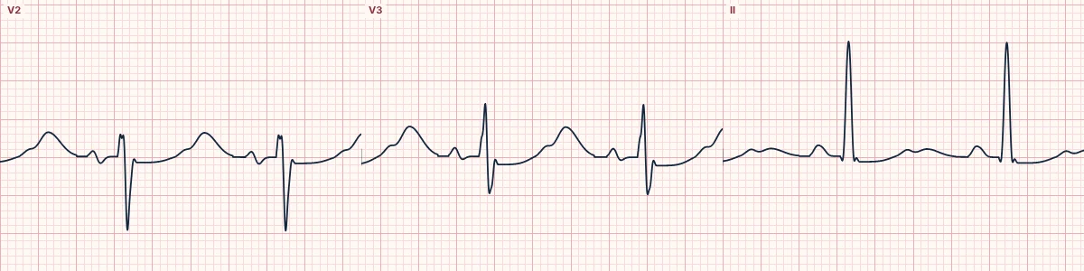
Example tracing, drawn by the lab's engine. Open the lab for a live, adjustable version.
At a glance
- T waves
- Flattened or inverted
- U waves
- Prominent, best in V2–V3; may exceed the T wave
- ST segment
- Mild depression
- QT
- Appears long because T and U merge (QU interval)
Figures
Still images drawn by the lab's engine at 25 mm/s and 10 mm/mV, with the finding labeled. Select a figure to enlarge it.
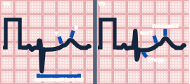
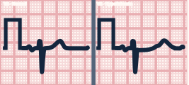
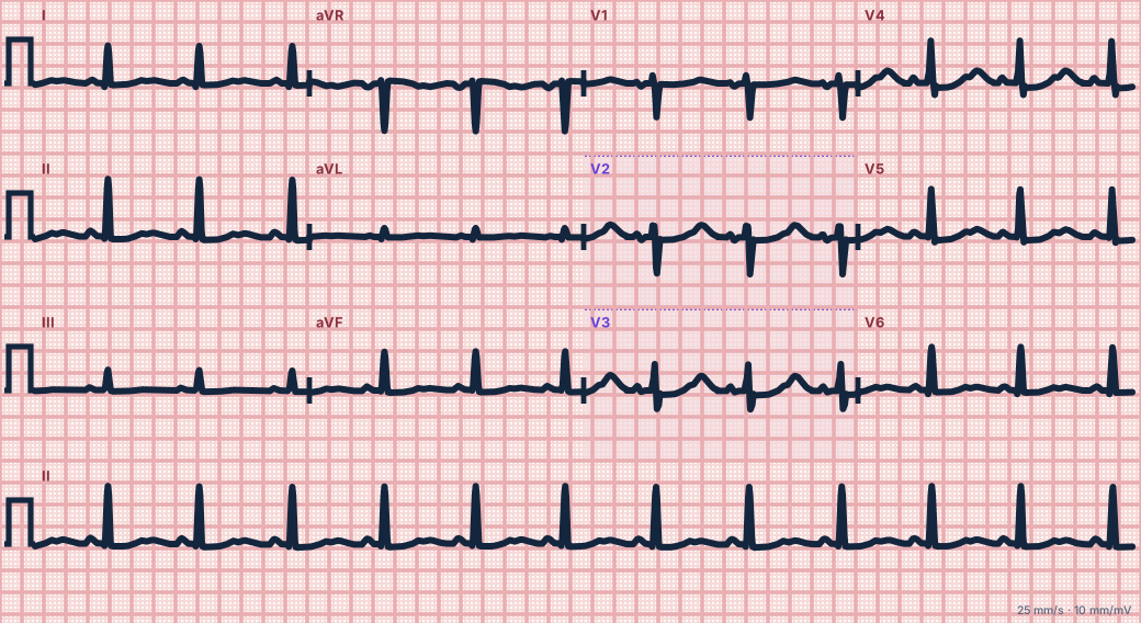
Scroll sideways to see the whole tracing, or select it to enlarge.
What the heart is doing
With less potassium outside the cells, the outward potassium current is weaker and repolarization is drawn out. The late part of repolarization shows up as a U wave.
Why it looks this way
This is hypokalemia because the T waves are flattened, the ST segment is mildly depressed and prominent U waves rise out of the T waves' tails.
Why it matters
Makes the ventricles irritable: PVCs, ventricular tachycardia and torsades de pointes, especially in patients on digoxin or with heart disease. Replace potassium, and magnesium with it.
What to look for
- In V2–V3, a U wave rising out of the end of the low T wave (T–U fusion), peaking about 0.1–0.15 s after it.
- The U wave is often taller than the T wave in V2–V3; T waves shrink as U waves grow.
- A seemingly very long QT is usually a QU interval: the T and U waves have fused.
Criteria
- T waves
- Flattened or inverted
- U waves
- Prominent, best in V2–V3; may exceed the T wave
- ST segment
- Mild depression
- QT
- Appears long because T and U merge (QU interval)
Causes
- Diuretics
- Vomiting and diarrhea
- Insulin or beta-agonist treatment
- Hypomagnesemia
- Excess aldosterone or steroids
Can be mistaken for
- Long QT syndrome — a single broad T wave, with no U wave rising out of its tail Compare
- Ischemia — ST depression without U waves, with symptoms
- Digoxin effect — scooped ST with a short QT Compare
Advanced Down to the ion channel
What the cells are doing
Pacemaker, atrial and ventricular action potentials, in step with the tracing above, with sodium, calcium and potassium moving through their channels.
Frequently asked questions
What are the ECG criteria for hypokalemia?
T waves: flattened or inverted. U waves: prominent, best in V2–V3; may exceed the T wave. ST segment: mild depression. QT: appears long because T and U merge (QU interval).
How is hypokalemia identified on an ECG?
This is hypokalemia because the T waves are flattened, the ST segment is mildly depressed and prominent U waves rise out of the T waves' tails.
What can hypokalemia be mistaken for?
Long QT syndrome: a single broad T wave, with no U wave rising out of its tail. Ischemia: ST depression without U waves, with symptoms. Digoxin effect: scooped ST with a short QT.
What causes hypokalemia?
Common causes: diuretics; vomiting and diarrhea; insulin or beta-agonist treatment; hypomagnesemia; and excess aldosterone or steroids.
Sources
This page does not list page-specific sources yet.
Every source the site cites, with links