
AV blocks
High-grade AV block
Two or more consecutive P waves fail to conduct, but some still get through. It sits between second-degree and complete block and behaves like the latter.
Last updated
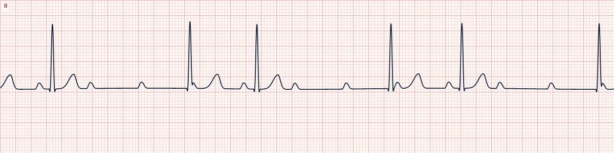
Example tracing, drawn by the lab's engine. Open the lab for a live, adjustable version.
At a glance
- Rate
- Atrial normal; ventricular slow, often < 40/min
- Rhythm
- Regular or irregular with escape beats
- P waves
- Regular; 3 or more for each conducted QRS
- PR interval
- Constant on the beats that conduct
Figures
Still images drawn by the lab's engine at 25 mm/s and 10 mm/mV, with the finding labeled. Select a figure to enlarge it.
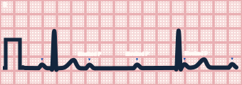
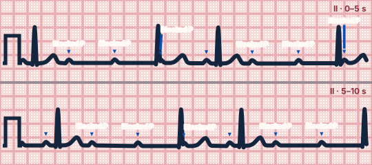
Scroll sideways to see the whole tracing, or select it to enlarge.
What the heart is doing
The AV node or His–Purkinje system fails to conduct most atrial impulses, passing only an occasional one. During long gaps, a junctional or ventricular escape focus may fire.
Why it looks this way
This is high-grade AV block because several consecutive P waves are not conducted, yet some QRS complexes still follow a P wave at a fixed PR.
Why it matters
A dangerous rhythm with a slow ventricular rate and a high risk of complete block or standstill. Symptomatic patients need urgent support and preparation for pacing; most need a pacemaker.
What to look for
- Count the P waves between QRS complexes: two or more in a row are not conducted.
- Some QRS complexes follow a P wave at a repeating, fixed PR, proving conduction still occurs.
- A QRS that appears without the usual PR is an escape beat.
Criteria
- Rate
- Atrial normal; ventricular slow, often < 40/min
- Rhythm
- Regular or irregular with escape beats
- P waves
- Regular; 3 or more for each conducted QRS
- PR interval
- Constant on the beats that conduct
- QRS
- Narrow or wide; escape beats may differ
Causes
- Myocardial infarction
- Degenerative conduction disease
- AV nodal blocking drug toxicity
- Hyperkalemia
- Myocarditis, Lyme disease
- After cardiac surgery
Can be mistaken for
- Third-degree AV block — no P wave ever conducts and the PR varies randomly
- 2:1 AV block — only one P wave is blocked at a time Compare
- Sinus bradycardia — one P wave per QRS Compare
Advanced Down to the ion channel
What the cells are doing
Pacemaker, atrial and ventricular action potentials, in step with the tracing above, with sodium, calcium and potassium moving through their channels.
Frequently asked questions
What are the ECG criteria for high-grade AV block?
Rate: atrial normal; ventricular slow, often < 40/min. Rhythm: regular or irregular with escape beats. P waves: regular; 3 or more for each conducted QRS. PR interval: constant on the beats that conduct. QRS: narrow or wide; escape beats may differ.
How is high-grade AV block identified on an ECG?
This is high-grade AV block because several consecutive P waves are not conducted, yet some QRS complexes still follow a P wave at a fixed PR.
What can high-grade AV block be mistaken for?
Third-degree AV block: no P wave ever conducts and the PR varies randomly. 2:1 AV block: only one P wave is blocked at a time. Sinus bradycardia: one P wave per QRS.
What causes high-grade AV block?
Common causes: myocardial infarction; degenerative conduction disease; AV nodal blocking drug toxicity; hyperkalemia; myocarditis, Lyme disease; and after cardiac surgery.
Sources
This page does not list page-specific sources yet.
Every source the site cites, with links