
AV blocks
First-degree AV block
Every atrial impulse reaches the ventricles, but conduction through the AV node is slow. The only finding is a PR interval longer than 200 ms.
Last updated
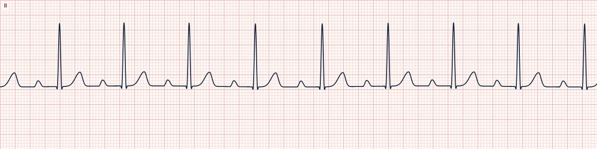
Example tracing, drawn by the lab's engine. Open the lab for a live, adjustable version.
At a glance
- Rate
- That of the underlying sinus rhythm
- Rhythm
- Regular
- P waves
- Normal; one before every QRS
- PR interval
- > 200 ms and constant
Figures
Still images drawn by the lab's engine at 25 mm/s and 10 mm/mV, with the finding labeled. Select a figure to enlarge it.
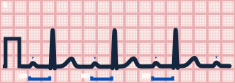
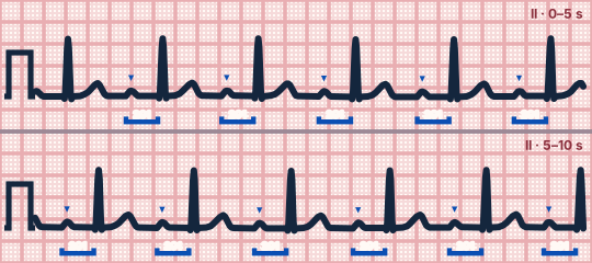
Scroll sideways to see the whole tracing, or select it to enlarge.
What the heart is doing
Impulses are delayed, usually within the AV node, but none is blocked. It is a conduction delay more than a true block: atria and ventricles stay linked 1:1.
Why it looks this way
This is first-degree AV block because every P wave conducts, with a constant PR longer than 200 ms.
Why it matters
Benign by itself and needs no treatment. Watch for progression when it is new in acute infarction, drug toxicity or infection, or when it occurs with bundle branch block.
What to look for
- Measure the PR from the start of the P wave to the start of the QRS: more than one large box.
- Confirm the PR is the same on every beat and that no P wave is left without a QRS.
- With a very long PR the P wave may sit on the preceding T wave.
Criteria
- Rate
- That of the underlying sinus rhythm
- Rhythm
- Regular
- P waves
- Normal; one before every QRS
- PR interval
- > 200 ms and constant
- QRS
- Narrow unless a bundle branch block coexists
Causes
- High vagal tone, athletic conditioning
- Beta-blockers, calcium-channel blockers, digoxin
- Inferior myocardial infarction
- Age-related conduction fibrosis
- Myocarditis, Lyme disease
- Hyperkalemia
Can be mistaken for
- Junctional rhythm — the P wave lost in the T wave can make the rhythm look P-less Compare
- 2:1 AV block — a second P wave is hidden in the T wave Compare
- Mobitz I — the PR changes from beat to beat and a QRS is dropped Compare
Advanced Down to the ion channel
What the cells are doing
Pacemaker, atrial and ventricular action potentials, in step with the tracing above, with sodium, calcium and potassium moving through their channels.
Frequently asked questions
What are the ECG criteria for first-degree AV block?
Rate: that of the underlying sinus rhythm. Rhythm: regular. P waves: normal; one before every QRS. PR interval: > 200 ms and constant. QRS: narrow unless a bundle branch block coexists.
How is first-degree AV block identified on an ECG?
This is first-degree AV block because every P wave conducts, with a constant PR longer than 200 ms.
What can first-degree AV block be mistaken for?
Junctional rhythm: the P wave lost in the T wave can make the rhythm look P-less. 2:1 AV block: a second P wave is hidden in the T wave. Mobitz I: the PR changes from beat to beat and a QRS is dropped.
What causes first-degree AV block?
Common causes: high vagal tone, athletic conditioning; beta-blockers, calcium-channel blockers, digoxin; inferior myocardial infarction; age-related conduction fibrosis; myocarditis, Lyme disease; and hyperkalemia.
Sources
This page does not list page-specific sources yet.
Every source the site cites, with links