
Waves, voltage and chambers
Draft — pending owner review
Katz–Wachtel phenomenon
Biventricular hypertrophy
Large biphasic RS complexes, over 50 mm from peak to trough, in the mid-chest leads V2–V4. It is the classic ECG sign of biventricular hypertrophy, most often in children with a ventricular septal defect.
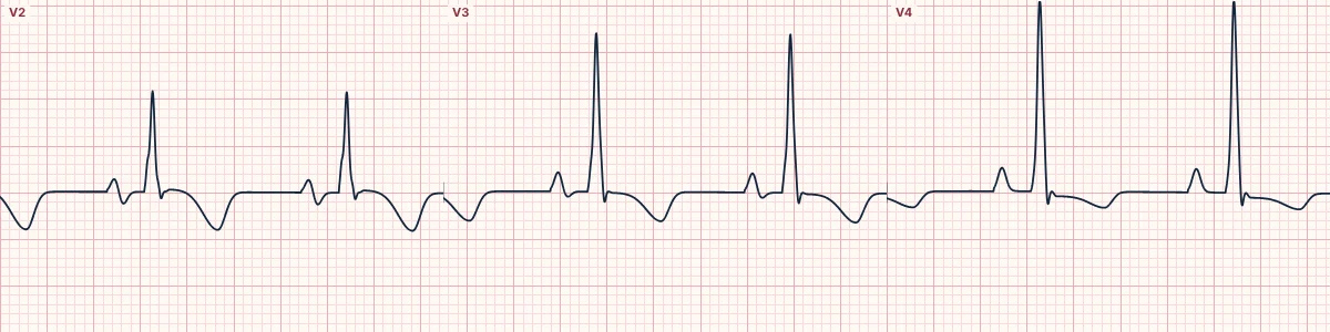
What it looks like
In V2, V3 or V4 the R and S waves are both large and roughly equal, so the complex is tall and biphasic. Together they often exceed 50 mm and can overlap the leads above and below on a printed page.
Why it happens
When both ventricles are hypertrophied, the mid-chest leads, which sit between them, see both large opposing forces: one writes the R wave and the other the S wave.
Other signs of biventricular hypertrophy
LVH voltage with right axis deviation; RVH features with a deep S in V1 and tall R in V5–V6; or a dominant R in V1 with left ventricular strain. Each is less specific than the Katz–Wachtel pattern.
About the demonstration
The tracing combines the lab's left and right ventricular hypertrophy models. It gives large mid-chest complexes but is a simplified illustration, not a reference for measuring.
How to read it
- Add the R and S amplitudes in V2–V4.
- Check the calibration first.
- Look for other features of right and left ventricular hypertrophy.
Pitfalls
- Thin chests in children and young adults give large voltages without hypertrophy.
- Overlapping leads on the printout are easily misread; re-record at half standard.
Frequently asked questions
Katz–Wachtel phenomenon: what is the key idea?
Large biphasic RS complexes, over 50 mm from peak to trough, in the mid-chest leads V2–V4. It is the classic ECG sign of biventricular hypertrophy, most often in children with a ventricular septal defect.
Katz–Wachtel phenomenon: what are the key numbers?
Leads: V2–V4; Amplitude: R + S > 50 mm; Classic setting: Ventricular septal defect in children; Tip: Record at half standard (5 mm/mV) to keep the complexes on the page.
Katz–Wachtel phenomenon: how do you apply it to an ECG?
Add the R and S amplitudes in V2–V4. Check the calibration first. Look for other features of right and left ventricular hypertrophy.
Katz–Wachtel phenomenon: what are the common pitfalls?
Thin chests in children and young adults give large voltages without hypertrophy. Overlapping leads on the printout are easily misread; re-record at half standard.