
Waves, voltage and chambers
Draft — pending owner review
LVH voltage criteria
Sokolow–Lyon, Cornell, Peguero–Lo Presti and Romhilt–Estes
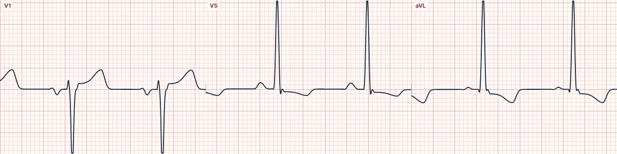
Several scoring rules turn QRS voltage into a diagnosis of left ventricular hypertrophy. They are specific but insensitive, so a normal score does not exclude LVH, and voltage alone misleads in young, thin people.
Why there are so many
Each rule captures the extra voltage of a thick left ventricle from a different angle. Most are specific, often above 85–90%, but each detects only a minority of hypertrophied hearts, and they perform differently with age, sex and body build.
Strain
Downsloping ST depression with asymmetric T-wave inversion in leads with tall R waves (I, aVL, V5–V6) is the repolarization pattern of LVH. It scores points in Romhilt–Estes, carries a worse prognosis, and can mimic or mask ischemia.
Supporting features
Left atrial enlargement, left axis deviation, a slightly wide QRS and a long R-wave peak time in V5–V6 support LVH when the voltage is borderline.
Who it misleads
Young, thin and athletic people often exceed voltage criteria without disease. Obesity, lung disease, pericardial effusion and LBBB lower the voltage or invalidate the rules.
How to read it
- Confirm standard calibration (10 mm/mV) before adding voltages.
- Work out at least Sokolow–Lyon and Cornell.
- Look for strain, left atrial enlargement and left axis deviation as support.
Pitfalls
- Half-standard calibration halves every voltage: double it before scoring.
- Do not apply adult criteria to children or adolescents.
Frequently asked questions
LVH voltage criteria: what is the key idea?
Several scoring rules turn QRS voltage into a diagnosis of left ventricular hypertrophy. They are specific but insensitive, so a normal score does not exclude LVH, and voltage alone misleads in young, thin people.
LVH voltage criteria: what are the key numbers?
Sokolow–Lyon: SV1 + RV5/V6 ≥ 35 mm; Cornell: RaVL + SV3 > 28 mm (men), > 20 mm (women); Peguero–Lo Presti: Deepest S + SV4 ≥ 28 mm (men), ≥ 23 mm (women); Romhilt–Estes: 4 points probable, ≥ 5 definite.
LVH voltage criteria: how do you apply it to an ECG?
Confirm standard calibration (10 mm/mV) before adding voltages. Work out at least Sokolow–Lyon and Cornell. Look for strain, left atrial enlargement and left axis deviation as support.
LVH voltage criteria: what are the common pitfalls?
Half-standard calibration halves every voltage: double it before scoring. Do not apply adult criteria to children or adolescents.