
Waves, voltage and chambers
Draft — pending owner review
Osborn (J) waves
A hump at the end of the QRS
A positive hump where the QRS meets the ST segment, most prominent in the inferior and lateral chest leads. It is the classic sign of hypothermia but also appears with high calcium and brain injury, and as a small notch in early repolarization.
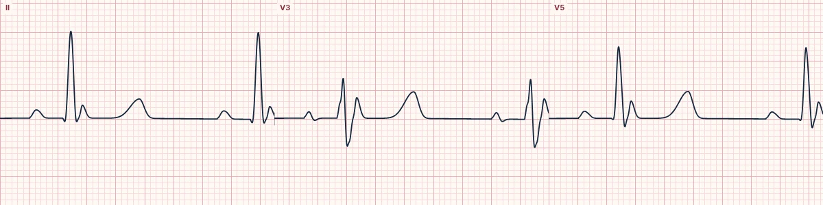
What it looks like
The QRS ends not in a sharp J point but in a dome or hump pointing the same way as the R wave, sometimes as tall as the R wave itself. It is often mistaken for an R′ or for ST elevation.
Hypothermia
The waves appear as the core temperature falls below about 32 °C and grow as it falls further, along with bradycardia, long PR, QRS and QT intervals, shivering artifact and atrial fibrillation. They shrink with rewarming.
Other causes
Severe hypercalcemia, raised intracranial pressure and subarachnoid hemorrhage, the period after cardiac arrest, and some idiopathic VF syndromes. A small notch at the end of the QRS is part of benign early repolarization.
Mechanism
A strong transient outward potassium current in the epicardium, but not the endocardium, opens a voltage gradient across the wall early in repolarization. Cold and calcium exaggerate it.
How to read it
- Look at the end of the QRS in the inferior and lateral leads for an extra positive deflection.
- Check the temperature and calcium.
- Watch for bradycardia and slow atrial fibrillation alongside.
Pitfalls
- Shivering artifact can hide or mimic J waves.
- Do not read the hump as the ST elevation of occlusion MI without other signs.
Frequently asked questions
Osborn (J) waves: what is the key idea?
A positive hump where the QRS meets the ST segment, most prominent in the inferior and lateral chest leads. It is the classic sign of hypothermia but also appears with high calcium and brain injury, and as a small notch in early repolarization.
Osborn (J) waves: what are the key numbers?
Best leads: II, III, aVF and V3–V6; Hypothermia: Usually below about 32 °C; Size: Grows as the temperature falls; Other names: J waves, camel-hump sign, hypothermic hump.
Osborn (J) waves: how do you apply it to an ECG?
Look at the end of the QRS in the inferior and lateral leads for an extra positive deflection. Check the temperature and calcium. Watch for bradycardia and slow atrial fibrillation alongside.
Osborn (J) waves: what are the common pitfalls?
Shivering artifact can hide or mimic J waves. Do not read the hump as the ST elevation of occlusion MI without other signs.