
Electrolytes
Hyperkalemia, moderate
As potassium rises further, conduction slows everywhere: the P wave flattens, the PR lengthens and the QRS begins to widen beneath peaked T waves.
Last updated
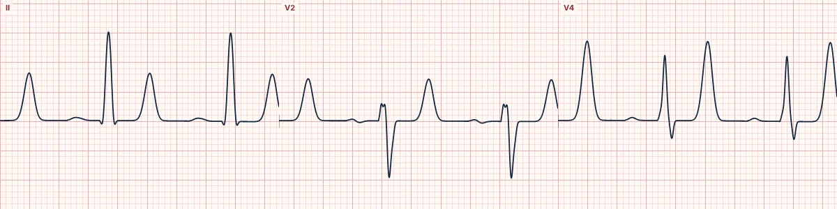
Example tracing, drawn by the lab's engine. Open the lab for a live, adjustable version.
At a glance
- P waves
- Flattened and widened
- PR interval
- Prolonged (> 200 ms)
- QRS
- Widened (≥ 120 ms)
- T waves
- Tall, peaked, symmetric
Figures
Still images drawn by the lab's engine at 25 mm/s and 10 mm/mV, with the finding labeled. Select a figure to enlarge it.
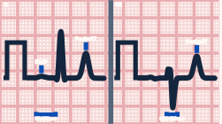
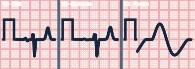
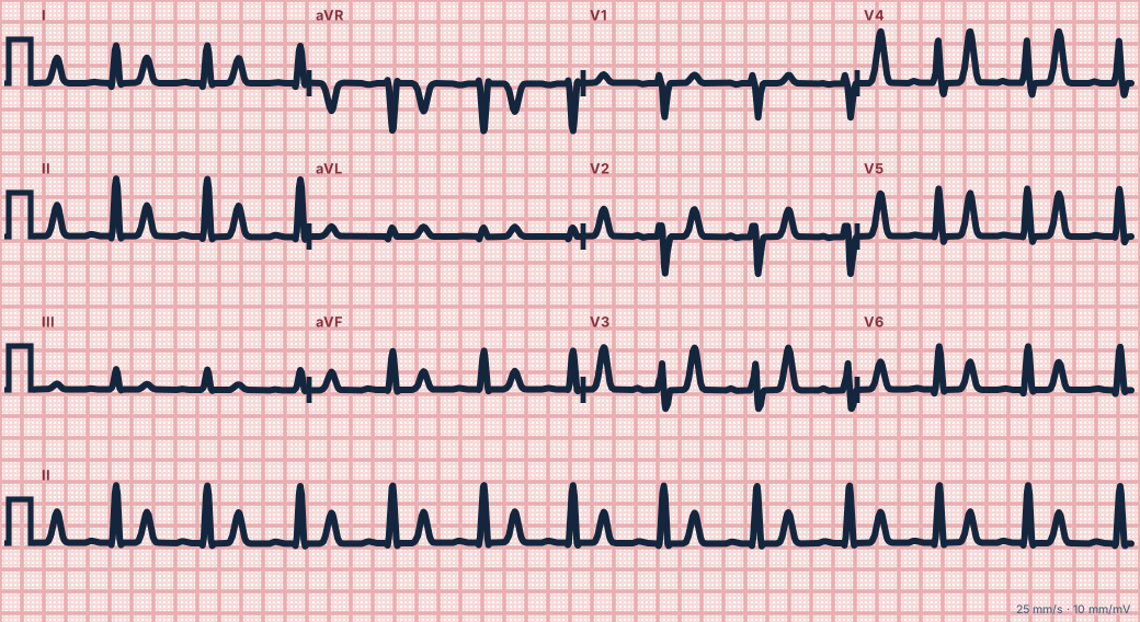
Scroll sideways to see the whole tracing, or select it to enlarge.
What the heart is doing
High potassium partly depolarizes resting cells and inactivates sodium channels. Conduction slows in the atria, AV node and ventricles together, with atrial muscle the most sensitive.
Why it looks this way
This is moderate hyperkalemia because flattened P waves, a long PR and a widened QRS accompany tall peaked T waves.
Why it matters
A medical emergency with a real risk of sudden bradycardia, heart block or ventricular arrhythmia. Give calcium to stabilize the heart, shift potassium into cells and arrange its removal.
What to look for
- P waves that are hard to find in a rhythm that is otherwise regular.
- A QRS that is uniformly broad without a typical bundle branch block pattern.
- Peaked T waves on top of a wide QRS: think potassium first.
Criteria
- P waves
- Flattened and widened
- PR interval
- Prolonged (> 200 ms)
- QRS
- Widened (≥ 120 ms)
- T waves
- Tall, peaked, symmetric
Causes
- Kidney failure or missed dialysis
- ACE inhibitors, potassium-sparing diuretics
- Acidosis, including diabetic ketoacidosis
- Tissue breakdown (crush injury, rhabdomyolysis, burns)
- Adrenal insufficiency
Can be mistaken for
- Bundle branch block — typical V1 and V6 patterns with normal P waves
- Sodium-channel blocker toxicity — wide QRS with a tall R in aVR, no peaked T waves Compare
- Junctional rhythm — P waves absent but the QRS is narrow Compare
Advanced Down to the ion channel
What the cells are doing
Pacemaker, atrial and ventricular action potentials, in step with the tracing above, with sodium, calcium and potassium moving through their channels.
Frequently asked questions
What are the ECG criteria for hyperkalemia, moderate?
P waves: flattened and widened. PR interval: prolonged (> 200 ms). QRS: widened (≥ 120 ms). T waves: tall, peaked, symmetric.
How is hyperkalemia, moderate identified on an ECG?
This is moderate hyperkalemia because flattened P waves, a long PR and a widened QRS accompany tall peaked T waves.
What can hyperkalemia, moderate be mistaken for?
Bundle branch block: typical V1 and V6 patterns with normal P waves. Sodium-channel blocker toxicity: wide QRS with a tall R in aVR, no peaked T waves. Junctional rhythm: P waves absent but the QRS is narrow.
What causes hyperkalemia, moderate?
Common causes: kidney failure or missed dialysis; ACE inhibitors, potassium-sparing diuretics; acidosis, including diabetic ketoacidosis; tissue breakdown (crush injury, rhabdomyolysis, burns); and adrenal insufficiency.
Practise it in the ACLS Lab
Sources
This page does not list page-specific sources yet.
Every source the site cites, with links