
Electrolytes
Hyperkalemia, mild (peaked T waves)
The earliest ECG sign of a raised blood potassium: tall, narrow, pointed T waves, while the P wave and QRS still look normal.
Last updated
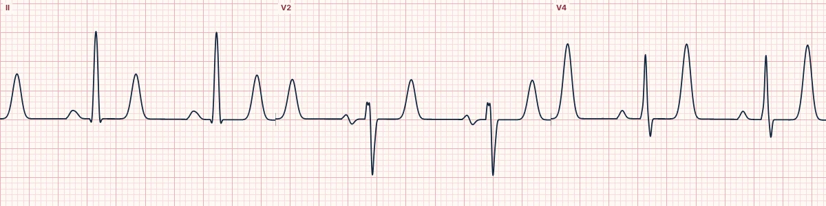
Example tracing, drawn by the lab's engine. Open the lab for a live, adjustable version.
At a glance
- T waves
- Tall, narrow, symmetric ('tented'), best in V2–V4
- QT
- Normal or short
- P waves
- Normal
- QRS
- Normal width
Figures
Still images drawn by the lab's engine at 25 mm/s and 10 mm/mV, with the finding labeled. Select a figure to enlarge it.
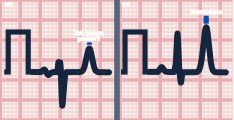
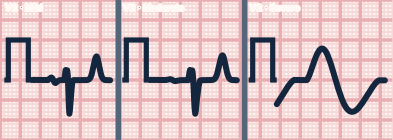
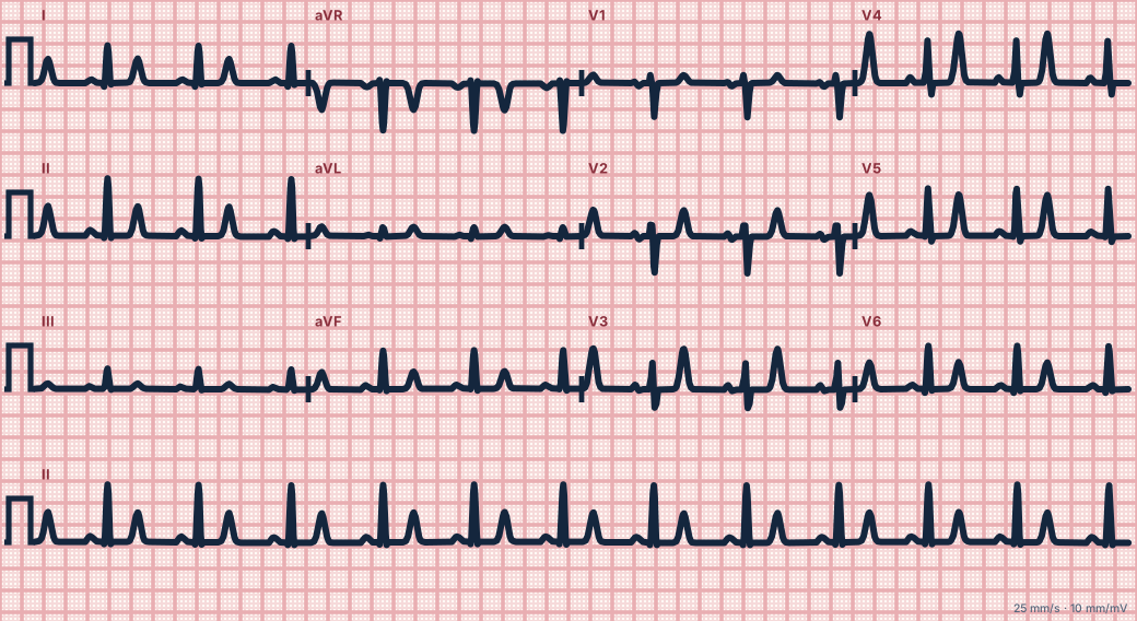
Scroll sideways to see the whole tracing, or select it to enlarge.
What the heart is doing
Extra potassium outside the cells speeds up repolarization. The T wave, which records repolarization, becomes taller, narrower and more symmetric before conduction itself is affected.
Why it looks this way
This is mild hyperkalemia because the T waves are tall, narrow and symmetric across many leads while the P waves and QRS remain normal.
Why it matters
The ECG tracks the potassium level poorly and can deteriorate quickly. Any change in an at-risk patient calls for urgent confirmation, continuous monitoring and treatment to protect the heart and lower potassium.
What to look for
- T waves with a narrow base and a sharp peak, as if pinched upward.
- The change is widespread, not confined to one coronary territory.
- Compare with an old ECG; a T taller than the R wave in several leads is suspicious.
Criteria
- T waves
- Tall, narrow, symmetric ('tented'), best in V2–V4
- QT
- Normal or short
- P waves
- Normal
- QRS
- Normal width
Causes
- Kidney failure or missed dialysis
- ACE inhibitors, potassium-sparing diuretics
- Acidosis, including diabetic ketoacidosis
- Tissue breakdown (crush injury, rhabdomyolysis, burns)
- Adrenal insufficiency
Can be mistaken for
- Hyperacute T waves of early STEMI — broad-based and limited to one territory
- Early repolarization — tall T waves with J-point elevation Compare
- de Winter pattern — tall T waves that begin from a depressed J point Compare
Advanced Down to the ion channel
What the cells are doing
Pacemaker, atrial and ventricular action potentials, in step with the tracing above, with sodium, calcium and potassium moving through their channels.
Frequently asked questions
What are the ECG criteria for hyperkalemia, mild (peaked T waves)?
T waves: tall, narrow, symmetric ('tented'), best in V2–V4. QT: normal or short. P waves: normal. QRS: normal width.
How is hyperkalemia, mild (peaked T waves) identified on an ECG?
This is mild hyperkalemia because the T waves are tall, narrow and symmetric across many leads while the P waves and QRS remain normal.
What can hyperkalemia, mild (peaked T waves) be mistaken for?
Hyperacute T waves of early STEMI: broad-based and limited to one territory. Early repolarization: tall T waves with J-point elevation. de Winter pattern: tall T waves that begin from a depressed J point.
What causes hyperkalemia, mild (peaked T waves)?
Common causes: kidney failure or missed dialysis; ACE inhibitors, potassium-sparing diuretics; acidosis, including diabetic ketoacidosis; tissue breakdown (crush injury, rhabdomyolysis, burns); and adrenal insufficiency.
Sources
This page does not list page-specific sources yet.
Every source the site cites, with links